Research - (2020) Advances in Dental Surgery
Comparative Evaluation of Canal Centering Ability of Three File Systems, Wave One, Wave One Gold and Protaper Gold File Systems-An In Vitro Study
Swathi UB, Sindhu Ramesh* and Pradeep S
*Correspondence: Sindhu Ramesh, Department of Conservative Dentistry and Endodontics, Saveetha Dental College, Saveetha Institute of Medical and Technical Sciences (SIMATS), Saveetha University, Chennai, India, Email:
Abstract
Introduction: Successful root canal therapy depends on effective debridement of the root canal by eliminating debris and microorganisms and shaping of the root canal system without deviating from the original anatomy. The ability to keep the instruments centered in curved canals and to deliver an accurate enlargement to the root canal without any unnecessary weakening to the root structure is crucial. Aim: This study aimed to compare the canal-centering ability of Protaper gold, WaveOne (WO), Wave one gold file systems using cone-beam computed tomography. Materials and Methods: In this in vitro study, forty extracted human single-rooted mandibular premolars were used. Pre instrumentation CBCT of all teeth were taken, canal curvatures were calculated, and the samples were randomly divided into two groups with ten samples in each group: Group 1–Protaper gold, Group 2–Wave one system, and Group 3- Wave one gold file system. Post instrumentation scans CBCT performed, analysed both the CBCT using DICOM software, to determine the canal-centering ability at 3 mm, 8 mm and 12 mm from the root apex. Statistical analysis: One-way anova and post hoc tests were used for statistical analysis in the present study. The mean and standard deviation values for canal-centering ratio was determined at the levels of 3, 8 and 12 mm between three groups, shaped with protaper gold, wave one and wave one gold file systems. Results: Using one way anova and post hoc, results were as follows: for canal-centering ability, Group 1 (Protaper gold), Group 2 (Wave one), Group 3 (Wave one gold) showed no statistically significant difference at 3 mm, 8 mm, and 12 mm (p>0.05) with slightly higher canal centering ability for the wave one file systems. Conclusion: Within the limitation of this study, Wave one file system has better canal-centering ability, maintains original canal curvature, and preserves more dentine as compared to wave one gold, protaper gold file systems.
Keywords
Canal centering, Cone-beam computed tomography, Root canal anatomy, Rotary files, Reciprocation files
Introduction
Successful root canal therapy depends on proper diagnosis, effective debridement of the root canal by eliminating debris and microorganisms and shaping of the root canal system without deviating from the original anatomy [1-4]. Ideally, during root canal preparation, the instruments should always conform to and retain the original shape of the canal. The ability to keep the instruments centered in curved canals and to deliver an accurate enlargement to the root canal without any unnecessary weakening to the root structure is crucial. A prepared root canal should have a continuously tapered funnel shape while maintaining the original outline form of the canal [15-18]. When curvatures are present, preparation becomes more difficult and there is a tendency for all preparation techniques to divert the prepared canal away from the original axis. Certain Factors that affect canal-centering ability are design of the instrument include cross section, taper, tip size, and flexibility.
Deviation from the original canal curvature can lead to, excessive and inappropriate dentin removal [19]. Straightening of the canal and creation of a ledge in the dentinal wall [20], a biochemical defect known as an elbow which forms the coronal to the ellipticalshaped apical seal [21], canals with hourglass appearance in cross-section that requires stripping, overpreparation that weakens the tooth, resulting in fracture of the root [19]. Ni- Ti endodontic instruments were introduced to facilitate instrumentation of curved canals. Ni- Ti instruments are superelastic and could flex far more than stainless steel instruments before exceeding their elastic limits [22-26].
Parameswaran et al. [27] Al omarii et al. [28] Coleman et al. [29] and Miglani et al. [30] reported transportation, zipping, and straightening of canals using stainless steel instruments. Several studies have confirmed that rotary Ni-Ti files maintain the original canal curvature better than stainless steel files [15,17,31]. The stainlesssteel files produce a larger extent of movement because of their hardness, which was shown to be 3-4 times harder than Ni-Ti alloys [8]. Carvalho reported that even after precurving and anticurvature filing, a small amount of transportation could be expected from stainless steel instruments [17].
WaveOne (WO) represents a single NiTi file system which is made up of a special NiTi alloy called M-Wire that is created by an innovative thermal treatment process. The benefits of this M-Wire NiTi alloy are increased flexibility and improved resistance to cyclic fatigue [32]. ProTaper Gold (PTG) and Wave One Gold (WOG) are relatively new canal preparation files resulting from advancement in NiTi systems. PTG files were developed with proprietary advanced metallurgy and have a progressively tapered design that is claimed by the manufacturer to enhance cutting efficiency and safety. PTG files have a convex triangular cross-section and progressive taper. This navigates challenging curves in the apical region of the canal. The file also has a feature of a shorter 11 mL handle for improved accessibility to the teeth.
WOG reciprocating single-file instrument system has a distinctive gold appearance. It has a unique parallelogram shaped cross-sectional design with one or two cutting edges depending on the location along the file. These edges significantly reduce torque, minimize screwing effect on the cutting efficiency, and allow better removal of debris. The distinctive gold appearance of the PTG and WOG files results from a unique heat treatment process that is applied after manufacturing of the files. The raw metal is repeatedly heated and cooled, giving it not only its gold color, but also considerably improved strength and flexibility [33].
Cone-beam computed tomography (CBCT) utilizes a cone-shaped X-ray beam and an area detector that captures a cylindrical volume of data in one acquisition, which is also used in the analysis of the root canal area, and parameters such as canal transportation, centering ratio, and the amount of root dentin removed by endodontic instruments [24,34,35]. Thus, the purpose of this study was to evaluate and compare the canal transportation ability, of wave one, wave one gold, protaper gold files systems using CBCT.
Materials and Methods
Forty freshly extracted human single-rooted mandibular premolar teeth for an orthodontic treatment purpose or for periodontal reasons extracted were collected for this study. Teeth without any previous endodontic treatment, fractures, resorptive defects, calcifications, or open apices were included for the study. They were cleaned of any residual tissue tags, rinsed under running water, and stored in 10% formalin solution. The presence of a single root and root canal in each tooth was confirmed on radiographs. To get the flat reference, the crowns were decoronated with a diamond disk (DFS, Germany) and a final dimension of 15-mm length was achieved for each tooth.
The root canal length was established by measuring the penetration of a size 10 K-file (Dentsply/Maillefer, Switzerland) until it reached the apical foramen and then subtracting 0.5 mm. Angle of curvature was assessed according to the criteria described by Schneider [22]. Teeth were radiographed using radiovisiography in buccolingual direction. A line was drawn parallel to the long axis of the canal. A second line was drawn from the apical foramen to intersect with the first at the point where the canal began to leave the long axis of the tooth. The acute angle thus formed was measured and the angle of curvature was thus determined. Further, only teeth with a degree of curvature ranging between 10° and 24° were included in the study. All teeth were scanned using CBCT before instrumentation. The exposure time was 3.0 s, operating at 75 kV and 2.0 mA. The images were stored in the computer’s hard disk for further comparison between pre- and postinstrumentation data using DICOM software. The teeth were then randomly divided into three groups of ten teeth each.
Groups (N=30)
Group 1- shaping done with protaper gold system (n=10).
Group 2-shaping done with wave one file system (n=10).
Group 3-shaping done with wave one gold file system (n=10) (Figures 1 to 5).
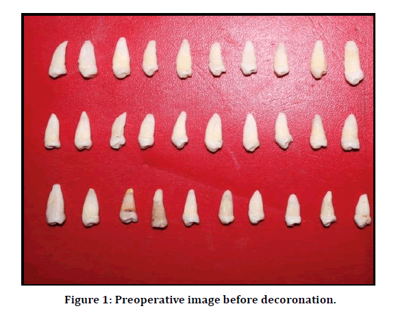
Figure 1: Preoperative image before decoronation.
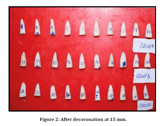
Figure 2: After decoronation at 15 mm.

Figure 3: Preoperative images of three groups.
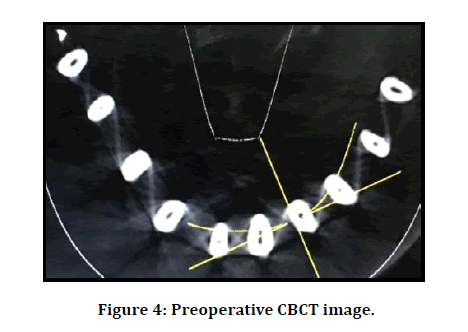
Figure 4: Preoperative CBCT image.
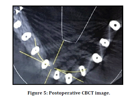
Figure 5: Postoperative CBCT image.
In all the three groups Canals were shaped using wave one rotary files, wave one gold files, protaper gold file having a size of 25 and a taper of 0.8 in a reciprocating, slow in‑and‑out pecking motion according to the manufacturer’s instructions. X‑Smart Plus endo motor (Dentsply Maillefer, Switzerland) was used. After each instrumentation, the canals were irrigated copiously with 3% sodium hypochlorite and the flutes of the instrument were cleaned. The final apical preparation size was 25 for the groups. Again, all the specimens were scanned by CBCT after instrumentation. Pre‑ and post‑instrumentation images were compared using DICOM software.
Canal‑centering ratio calculation
The canal‑centering ratio was calculated according to the following ratio:
(a1−a2)/(b1−b2) or (b1−b2)/(a1−a2)
The canal‑centering ratio is the difference between the instrumented and no instrumented canals, which measures the ability of an instrument to stay centered. If the numbers are not equal, the lower figure is considered as the numerator and a result of “1” indicates perfect canal‑centering capacity and, the closer the result to 0, the worse the ability of the instrument to keep itself in the canal’s central axis where a1 is the shortest distance from the mesial edge of the root to the mesial edge of the uninstrumented canal, b1 is shortest distance from the distal edge of the root to the distal edge of the uninstrumented canal, a2 is the shortest distance from the mesial edge of the root to the mesial edge of the instrumented canal, and b2 is the shortest distance from the distal edge of the root to the distal edge of the instrumented canal.
Results and Discussion
One-way anova and post hoc was used for statistical analysis with 0.05 level of significance. The mean and standard deviation values for canal‑centering ratio was determined at the levels of 3, 8 and 12 mm between three groups. However, for Wave one showed slightly better canal centering ability compared with the other two file systems (Tables 1 and 2) (Figures 6-8).
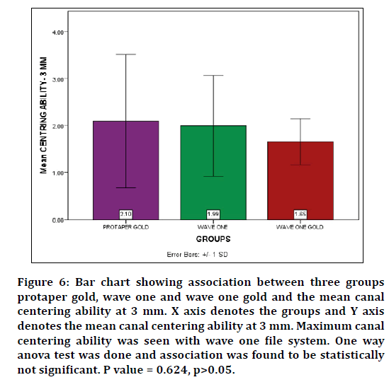
Figure 6: Bar chart showing association between three groups protaper gold, wave one and wave one gold and the mean canal centering ability at 3 mm. X axis denotes the groups and Y axis denotes the mean canal centering ability at 3 mm. Maximum canal centering ability was seen with wave one file system. One way anova test was done and association was found to be statistically not significant. P value = 0.624, p>0.05.
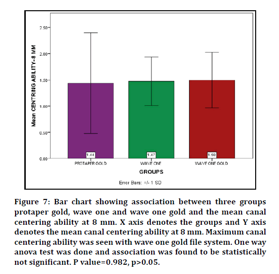
Figure 7: Bar chart showing association between three groups protaper gold, wave one and wave one gold and the mean canal centering ability at 8 mm. X axis denotes the groups and Y axis denotes the mean canal centering ability at 8 mm. Maximum canal centering ability was seen with wave one gold file system. One way anova test was done and association was found to be statistically not significant. P value=0.982, p>0.05.
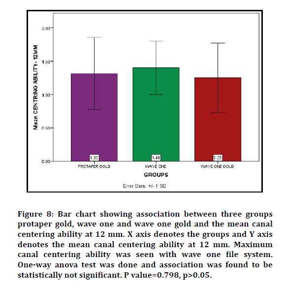
Figure 8: Bar chart showing association between three groups protaper gold, wave one and wave one gold and the mean canal centering ability at 12 mm. X axis denotes the groups and Y axis denotes the mean canal centering ability at 12 mm. Maximum canal centering ability was seen with wave one file system. One-way anova test was done and association was found to be statistically not significant. P value=0.798, p>0.05.
| Groups | Centring ability- 3 MM | Centring ability-8 MM | Centring ability-12MM | |
|---|---|---|---|---|
| PROTAPER GOLD | Mean | 2.1 | 1.437 | 1.3165 |
| N | 10 | 10 | 10 | |
| Std. Deviation | 1.42127 | 0.96051 | 0.54115 | |
| WAVE ONE | Mean | 1.988 | 1.473 | 1.401 |
| N | 10 | 10 | 10 | |
| Std. Deviation | 1.07621 | 0.46433 | 0.40336 | |
| WAVE ONE GOLD | Mean | 1.651 | 1.496 | 1.253 |
| N | 10 | 10 | 10 | |
| Std. Deviation | 0.48734 | 0.5333 | 0.52235 | |
| Total | Mean | 1.913 | 1.4687 | 1.3235 |
| N | 30 | 30 | 30 | |
| Std. Deviation | 1.04772 | 0.66491 | 0.47943 |
Table 1: Showing mean, standard deviation of all the three groups protaper gold, wave one gold and wave one file systems.
| ANOVA | ||||||
|---|---|---|---|---|---|---|
| Sum of Squares | df | Mean Square | F | Sig. | ||
| Centring ability-3 MM | Between groups | 1.092 | 2 | 0.546 | 0.48 | 0.624 |
| Within groups | 30.741 | 27 | 1.139 | |||
| Total | 31.834 | 29 | ||||
| Centring ability-8 MM | Between Groups | 0.018 | 2 | 0.009 | 0.019 | 0.982 |
| Within Groups | 12.803 | 27 | 0.474 | |||
| Total | 12.821 | 29 | ||||
| Centring ability-12 MM | Between Groups | 0.11 | 2 | 0.055 | 0.227 | 0.798 |
| Within Groups | 6.556 | 27 | 0.243 | |||
| Total | 6.666 | 29 | ||||
Table 2: Results of one way anova statistical test showing no statistically significant difference between the three groups way one,way one gold and protaper gold file systems.
None of the three systems evaluated had perfect canal‑centering ability. In the present study, we have utilized wave one, wave one gold, protaper gold NiTi file systems and the final apical preparation size was 25 for all the groups. Wave one NiTi single‑file reciprocating system has different cross‑sectional designs over the entire length of the working part. In the tip region, the cross section of the file presents radial lands, and in the middle portion of the working length and near the shaft, the cross‑sectional design changes from a modified triangular convex cross section with radial lands to that of a neutral rake angle with a triangular convex cross section [25,27,28]. Only one single‑shaping file is required to provide the canal with an adequate size and taper. Purpose of this design is to eliminate threading and binding of the instrument in continuous rotation. Although there is literature on the reduction of fatigue and extended life span of the instrument, there is a requirement of investigations regarding canal‑shaping ability of single‑file systems [29].
ProTaper Gold (PTG) and WaveOne Gold (WOG) are relatively new canal preparation files resulting from advancement in NiTi systems. PTG files were developed with proprietary advanced metallurgy and have a progressively tapered design that is claimed by the manufacturer to enhance cutting efficiency and safety. PTG files have a convex triangular cross-section and progressive taper. This navigates challenging curves in the apical region of the canal. The file also has a feature of a shorter 11mL handle for improved accessibility to the teeth.
WOG reciprocating single-file instrument system has a distinctive gold appearance. It has a unique parallelogram shaped cross-sectional design with one or two cutting edges depending on the location along the file. These edges significantly reduce torque, minimize screwing effect on the cutting efficiency, and allow better removal of debris.
The distinctive gold appearance of the PTG and WOG files is the result of a unique heat treatment process applied after manufacture. The raw metal is repeatedly heated and cooled, giving it not only its gold color, but also considerably improved strength and flexibility [33].
There is no statistical significance with the canal centering ability of wave one, wave one gold, protaper gold file systems. However, the Wave one file system has a slightly better canal-centering ability, maintains original canal curvature and preserves more dentine in comparison to wave one gold, protaper gold file systems.
Conclusion
Within the limitation of this study , all the NiTi rotary systems tested performed similarly with regard to canal centering ability and were able to maintain the original canal curvature and canal centering ability in mandibular premolars it was found that Wave one file system produced a slightly better canal centering ability, remained centered and respected the original canal anatomy in comparison with protaper gold and wave one gold file systems with an apical instrumentation diameter of #25.Wave one file system has a better canal centering ability compared to wave one gold and protaper taper gold file systems.
Data Availability
The experimental data used to support the findings of this study are included within the article.
Conflict of Interest
The authors of this study declare that there are no conflicts of interest with regard to the publication of this paper.
Acknowledgement
With Sincere gratitude, we acknowledge the staff members of the department of Conservative Dentistry and Endodontics, Saveetha Dental College and study participants for their extended support towards the completion of research.
References
- Govindaraju L, Neelakantan P, Gutmann JL. Effect of root canal irrigating solutions on the compressive strength of tricalcium silicate cements. Clin Oral Investigations 2017; 21:567–571.
- Azeem RA, Sureshbabu NM. Clinical performance of direct versus indirect composite restorations in posterior teeth: A systematic review. J Conservative Dent 2018; 21:2–9.
- Jenarthanan S, Subbarao C. Comparative evaluation of the efficacy of diclofenac sodium administered using different delivery routes in the management of endodontic pain: A randomized controlled clinical trial. J Conservative Dent 2018; 21:297–301.
- Manohar MP, Sharma S. A survey of the knowledge, attitude, and awareness about the principal choice of intracanal medicaments among the general dental practitioners and nonendodontic specialists. Indian J Dent Res 2018; 29:716–720.
- Nandakumar M, Nasim I. Comparative evaluation of grape seed and cranberry extracts in preventing enamel erosion: An optical emission spectrometric analysis. J Conservative Dent 2018; 21:516–520.
- Teja KV, Ramesh S, Priya V. Regulation of matrix metalloproteinase-3 gene expression in inflammation: A molecular study. J Conservative Dent 2018; 21:592–596.
- Khandelwal A, Palanivelu A. Correlation between dental caries and salivary albumin in adult population in Chennai: An In Vivo study. Brazilian Dent Sci 2019; 22:228–233.
- Malli Sureshbabu N, Selvarasu K, Nandakumar M, et al. Concentrated growth factors as an ingenious biomaterial in regeneration of bony defects after periapical surgery: A report of two cases. Case Reports Dent 2019; 7046203.
- Poorni S, Srinivasan MR, Nivedhitha MS. Probiotic strains in caries prevention: A systematic review. J Conservative Dent 2019; 22:123–128.
- Rajakeerthi R, Ms N. Natural product as the storage medium for an avulsed tooth–A systematic review. Cumhuriyet Dent J 2019; 22:249–256.
- Rajendran R, Kunjusankaran RN, Sandhya R, et al. Comparative evaluation of remineralizing potential of a paste containing bioactive glass and a topical cream containing casein phosphopeptide-Amorphous calcium phosphate: An in Vitro study. Pesquisa Brasileira Odontopediatria Clin Integrada 2019; 19:1–10.
- Ramarao S, Sathyanarayanan U. CRA Grid-A preliminary development and calibration of a paper-based objectivization of caries risk assessment in undergraduate dental education. J Conservative Dent 2019; 22:185–190.
- Siddique R, Sureshbabu NM, Somasundaram J, et al. Qualitative and quantitative analysis of precipitate formation following interaction of chlorhexidine with sodium hypochlorite, neem, and tulsi. J Conservative Dent 2019; 22:40–47.
- Siddique R, Nivedhitha MS, Jacob B. Quantitative analysis for detection of toxic elements in various irrigants, their combination (precipitate), and para-chloroaniline: An inductively coupled plasma mass spectrometry study. J Conservative Dent 2019; 22:344–350.
- Schilder H. Cleaning and shaping the root canal. Dent Clin North Am 1974; 18:269–296.
- Tsesis I, Amdor B, Tamse A, et al. The effect of maintaining apical patency on canal transportation. Int Endodont J 2008; 41:431–435.
- Kandaswamy D, Venkateshbabu N, Porkodi I, et al. Canal-centering ability: An endodontic challenge. J Conservative Dent 2009; 12:3–9.
- Teja KV, Ramesh S. Shape optimal and clean more. Saudi Endodont J 2019; 9:235.
- Gundappa M, Bansal R, Khoriya S, et al. Root canal centering ability of rotary cutting nickel titanium instruments: A meta-analysis. J Conservative Dent 2014; 17:504–509.
- Berutti E, Paolino DS, Chiandussi G, et al. Root canal anatomy preservation of WaveOne reciprocating files with or without glide path. J Endodont 2012; 38:101–104.
- Lim YJ, Park SJ, Kim HC, et al. Comparison of the centering ability of Wave\textperiodcentered One and Reciproc nickel-titanium instruments in simulated curved canals. Restorative Dent Endodont 2013; 38:21–25.
- Schneider SW. A comparison of canal preparations in straight and curved root canals. Oral Surg Oral Med Oral Pathol 1971; 32:271–275.
- Gambill JM, Alder M, del Rio CE. Comparison of nickel-titanium and stainless steel hand-file instrumentation using computed tomography. J Endodont 1996; 22:369–375.
- Maitin N, Arunagiri D, Brave D, et al. An ex vivo comparative analysis on shaping ability of four NiTi rotary endodontic instruments using spiral computed tomography. J Conservative Dent 2013; 16:219–223.
- Jain A, Asrani H, Singhal AC, et al. Comparative evaluation of canal transportation, centering ability, and remaining dentin thickness between WaveOne and ProTaper rotary by using cone beam computed tomography: An in vitro study. J Conservative Dent 2016; 19:440–444.
- Siddique R, Nivedhitha MS. Effectiveness of rotary and reciprocating systems on microbial reduction: A systematic review. J Conservative Dent 2019; 22:114–122.
- Arora A, Taneja S, Kumar M. Comparative evaluation of shaping ability of different rotary NiTi instruments in curved canals using CBCT. J Conservative Dent 2014; 17:35–39.
- Navós BV, Hoppe CB, Mestieri LB, et al. Centering and transportation: In vitro evaluation of continuous and reciprocating systems in curved root canals. J Conservative Dent 2016; 19:478–481.
- You SY, Bae KS, Baek SH, et al. Lifespan of one nickel-titanium rotary file with reciprocating motion in curved root canals. J Endodont 2010; 36:1991–1994.
- Hashem AA, Ghoneim AG, Lutfy RA, et al. Geometric analysis of root canals prepared by four rotary NiTi shaping systems. J Endodont 2012; 38:996–1000.
- Dhingra A, et al. Comparative evaluation of the canal curvature modifications after instrumentation with one shape rotary and wave one reciprocating files. J Conservative Dent 2014; 17:138–141.
- Lim YJ, Park SJ, Kim HC, et al. Comparison of the centering ability of Wave One and Reciproc nickel-titanium instruments in simulated curved canals. Restorative Dent Endodont 2013; 21.
- Madani ZS, Haddadi A, Haghanifar S, et al. Cone-beam computed tomography for evaluation of apical transportation in root canals prepared by two rotary systems. Iranian Endodont J Iranian Center Endodont Res 2014; 9:109.
- https://pdfs.semanticscholar.org/201e/e83f013736e5666a9d294862d47aaac21637.pdf.
- Janani K, Sandhya R. (2019) A survey on skills for cone beam computed tomography interpretation among endodontists for endodontic treatment procedure. Indian J Dent Res 2019; 30: 834–838.
Author Info
Swathi UB, Sindhu Ramesh* and Pradeep S
Department of Conservative Dentistry and Endodontics, Saveetha Dental College, Saveetha Institute of Medical and Technical Sciences (SIMATS), Saveetha University, Chennai, IndiaCitation: Swathi UB, Sindhu Ramesh, Pradeep S, Comparative Evaluation of Canal Centering Ability of Three File Systems, Wave One, Wave One Gold and Protaper Gold File Systems-An In Vitro Study, J Res Med Dent Sci, 2020, 8 (7): 447-453.
Received: 23-Sep-2020 Accepted: 10-Nov-2020 Published: 17-Nov-2020
