Case Report - (2022) Volume 10, Issue 5
Closed Reduction of Nasal Bone Fracture-A Case Series
Nithya Balasubramanian* and Rajasekar MK
*Correspondence: Nithya Balasubramanian, Department of ENT, Sree Balaji Medical College and Hospital, Chrompet, Chennai, India, Email:
Abstract
The most common type of facial bone fracture is a nasal bone fracture, which accounts for 40% to 50% of all instances. Nasal bone fractures can occur as a result of falls, physical assaults, car accidents, and blunt face injuries. They can occur on their own or in combination with other soft tissue and bone injuries to the face. Because of the protrusion of the nasal bones and its central location on the face, the nose is sensitive to harm. Nasal fractures are more common in males than in females. While nasal fractures are the most prevalent type of facial fracture, zygomatic-orbital complex fractures and skull base fractures are also possible.
Keywords
Facial bone, Nasal bone, HaemostasisIntroduction
Nasal bone fractures are linked to facial deformity and can cause airway obstruction. Deformity can be minimised and functional impairment can be avoided with early intervention. For easy, non-comminute fractures, a closed approach may be the best option. External digital pressure is applied over the fracture site with an instrument inserted into the nasal cavity. The bone is moved between the digit and the elevator until it is in the correct position. An Asch's and Walsham's forceps are used to realign the displaced nasal septum. To protect the nasal bone, an external splint is applied [1,2].
Case Presentation
A total of 25 cases of nasal bone fractures were treated by closed reduction. Patients presented with history of trauma to the nose mostly due to road traffic accidents, assault or direct blow to the face. Most patients came with complaints of nose bleed at the time of trauma and swelling at the site of fracture.
Physical examination showed an external deformity and depression on the nose with a deviated nasal septum to the right. X-Ray of the nasal bone was done which showed fracture of the nasal bone.
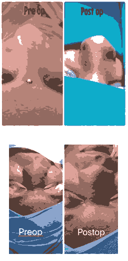
Under general anaesthesia, the patients had a closed reduction of the nasal fracture. Asch and Walsham forceps were used to decrease the nasal bone fracture. The nasal bone, which had been depressed, had been raised. The patient's haemostasis was obtained, and an external splint was applied. In the post-operative period, there were no significant issues.

Figure 1: Pre OP

Figure 2: Post OP

Figure 3:Pre OP and Post OP
Results and Discussion
A bony and cartilaginous structure supports the nose. The bony structure is made up of bilaterally paired nasal bones and the frontal process of the maxilla. The upper and lower lateral cartilages, as well as the septum, are cartilaginous structures. Both of these structures are prone to breaking [3,4].
Nasal bleeds are often linked to nasal bone fractures. The ophthalmic artery (a branch of the internal carotid artery) supplies the nose, splitting into anterior and posterior ethmoidal arteries. The external carotid artery also supplies it, as do the facial and internal maxillary arteries. Any trauma to the nose may cause bleeding from Keisselbach's plexus. The Kiesselbach plexus is created by the anastomosis of the following arteries on the anteroinferior nasal septum [5].
A branch of the ophthalmic artery, the anterior ethmoidal artery, a branch of the maxillary artery, the sphenopalatine artery, a branch of the maxillary artery, the larger palatine artery. The superior labial artery which is a facial artery branch [6].
Classification of nasal bone fractures
Nasal bone fractures are graded on a scale that depicts the severity of the injury. Low-velocity trauma is the most common cause of an isolated nasal fracture.
Classification: Nasal bone fractures were classified into six types:
Type I: Simple without displacement
Type II: Simple with displacement/without telescoping
IIA: Unilateral
IIAs: Unilateral with septal fracture
IIB: Bilateral
IIBs: Bilateral with septal fracture
Type III: Comminuted with telescoping or depression
The history of the injury should include the mechanism of injury, direction and history of previous surgeries Any loss of consciousness should be noted and vital should be checked prior to further management [7,8].
Physical examination and imaging
A general examination is always performed to rule out severe, life-threatening conditions.
Imaging for isolated nasal fractures is rarely needed. CT scans are performed for suspected head injuries, basal skull fractures or complex facial injuries.
Conclusion
Closed reduction is the best option for simple, noncomminuted fractures, with a success rate of 60% to 90%. To reduce depressed nasal bone segments, digital pressure and an elevator can be employed. Asch's and Walsham's forceps can be placed into the nasal cavity and rotated laterally to outfracture the bones. The laterally displaced parts of the bony pyramid will be digitally manipulated by force acting in the opposite direction. The nasal septum should be thoroughly checked, and if necessary, the septal base should be adjusted. Patients should be aware that a subsequent septorhinoplasty may be required, as reoperation rates range from 9% to 17%. Patients should wear a nasal splint for seven days after surgery. It not only helps to keep bones in place, but it also serves as a reminder to the patient and people around them to be cautious because the bones can easily displace again. Internal splints are not necessary in most closed reductions, but they have been employed in comminuted fractures, septal dislocations, and inwardly collapsing nasal bones.
References
- Kim KS, Lee HG, Shin JH, et al. Trend Analysis of Nasal Bone Fracture. Arch Craniofac Surg 2018; 19:270-227
[crossref] [Googlescholar] [Indexed]
- Swenson DM, Yard EE, Collins CL, et al. Epidemiology of Us High School Sports-Related Fractures, 2005-2009. Clin J Sport Med 2010; 20:293-299
[crossref] [Googlescholar] [Indexed]
- Hwang K, Jung JS, Kim H, et al. Diagnostic Performance of Plain Film, Ultrasonography and Computed Tomography in Nasal Bone Fractures: A Systematic Review. Plast Surg 2018; 26:286-292
[crossref] [Googlescholar] [Indexed]
- Atighechi S, Karimi G. Serial Nasal Bone Reduction: A New Approach to the Management of Nasal Bone Fracture.J Craniofac Surg 2009; 20:49-52
[crossref] [Googlescholar] [Indexed]
- Khwaja S, Pahade AV, Luff D, et al. Nasal Fracture Reduction: Local Versus General Anaesthesia. Rhinol 2007; 45:83-88
[Googlescholar] [Indexed]
- Lu GN, Humphrey CD, Kriet JD, et al. Correction of Nasal Fractures.Facial Plast Surg Clin North Am 2017; 25:537-546
[crossref] [Googlescholar] [Indexed]
- Li K, Moubayed SP, Spataro E, et al. Risk Factors for Corrective Septorhinoplasty Associated With Initial Treatment of Isolated Nasal Fracture. JAMA Facial Plast Surg 2018; 20:460-467
[crossref] [Googlescholar] [Indexed]
- Higuera S, Lee EI, Cole P, et al. Nasal Trauma and the Deviated Nose. Plast Reconstr Surg 2007; 120:64S-75S
[crossref] [Googlescholar] [Indexed]
Author Info
Nithya Balasubramanian* and Rajasekar MK
Department of ENT, Sree Balaji Medical College and Hospital, Chrompet, Chennai, IndiaCitation: Nithya Balasubramanian, Rajasekar MK, Closed Reduction of Nasal Bone Fracture-a Case Series, J Research Med Dent Sci, 2022, 10 (5): 7-9.
Received: 21-Feb-2022, Manuscript No. 41489; , Pre QC No. 41489; Editor assigned: 23-Feb-2022, Pre QC No. 41489; Reviewed: 09-Mar-2022, QC No. 41489; Revised: 22-Apr-2022, Manuscript No. 41489; Published: 02-May-2022
