Image Article - (2022) Volume 10, Issue 3
Clinical Image: CT Findings in severe cases of COVID-19
Ravi Bastakoti1, Adarsh Jha2*, Rabin Raj Singh1, Trinetra Kumar Karna1, Kshitij Giri1, Sunil Regmi1 and Atul Dwivedi3
*Correspondence: Adarsh Jha, Vedanta Hospital, India, Email:
Abstract
Chest CT has a potential role in the diagnosis, detection of complications, and prognostication of coronavirus disease 2019 (COVID-19). Implementation of appropriate precautionary safety measures, chest CT protocol optimization, and a standardized reporting system based on the pulmonary findings in this disease will enhance the clinical utility of chest CT. However, chest CT examinations may lead to both false-negative and false-positive results. Furthermore, the added value of chest CT in diagnostic decision making is dependent on several dynamic variables, most notably available resources (real-time reverse transcription–polymerase chain reaction [RT-PCR] tests, personal protective equipment, CT scanners, hospital and radiology personnel availability, and isolation room capacity) and the prevalence of both COVID-19 and other diseases with overlapping manifestations at chest CT.
Keywords
Ground glass appearance, Crazy paving pattern, Covid-19
Introduction
Multiple number of case of unexplained pneumonia have been reported in Wuhan since 2019 December .As the disease spread across the globe and infected large number of people .A new type of corona virus , which has been detected as novel corona virus 2019 or COVID -19 in patients Respiratory secretions and finally identified as the source of infection [1]. CT was used as a means of rapid diagnosis and disease progression and prognosis in Hubei, China , with lower mortality rate than other parts of the world . Widespread Use of CT is also advocated as the tool of quick diagnosis and control measure towards COVID -19 pandemic [2-4]. While other including the Fleischner society, recommend the use of CT in selected clinical settings [5,6]. Here we presented 5 severe cases of covid- 19 along with their characteristic CT findings (Figures 1 to Figure 5).

Figure 1. Severe type: A 48 - year- old male was admitted to the hospital with cough and fever for half a month. CT scan showed multiple ground glass opacity and consolidation shadows in both lungs with” crazy paving pattern " and" air bronchogram sign.

Figure 2. Severe type. A 66-year-old make was admitted to the hospital with cough and fever for a week. CT scan showed multiple ground glass opacity and consolidation shadows in both lungs, which were widely distributed in various lobed, especially sub-pleural.

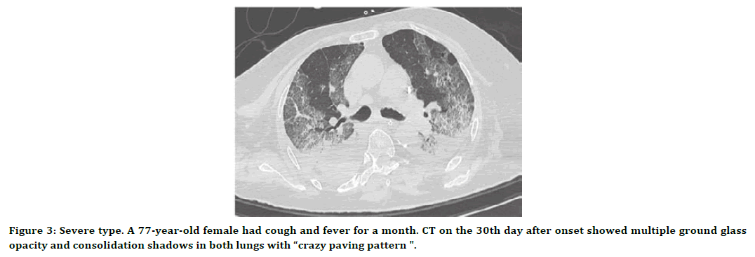
Figure 3. Severe type. A 77-year-old female had cough and fever for a month. CT on the 30th day after onset showed multiple ground glass opacity and consolidation shadows in both lungs with “crazy paving pattern ".

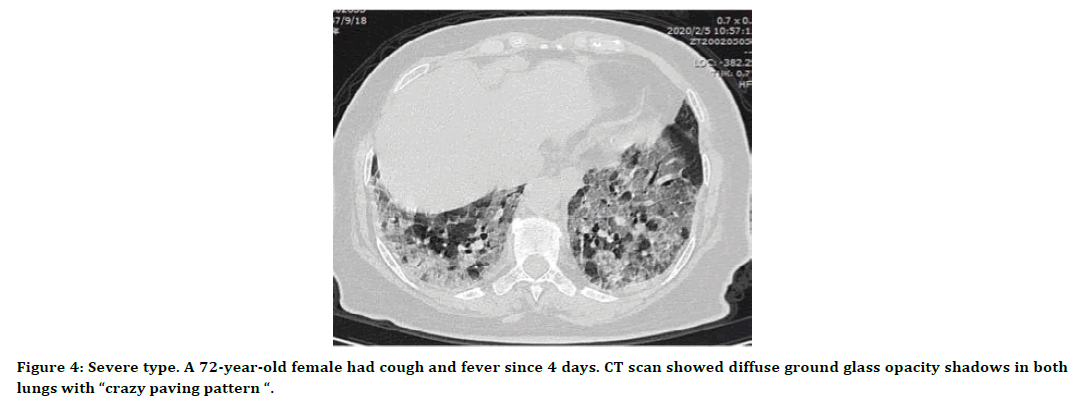
Figure 4. Severe type. A 72-year-old female had cough and fever since 4 days. CT scan showed diffuse ground glass opacity shadows in both lungs with “crazy paving pattern “.

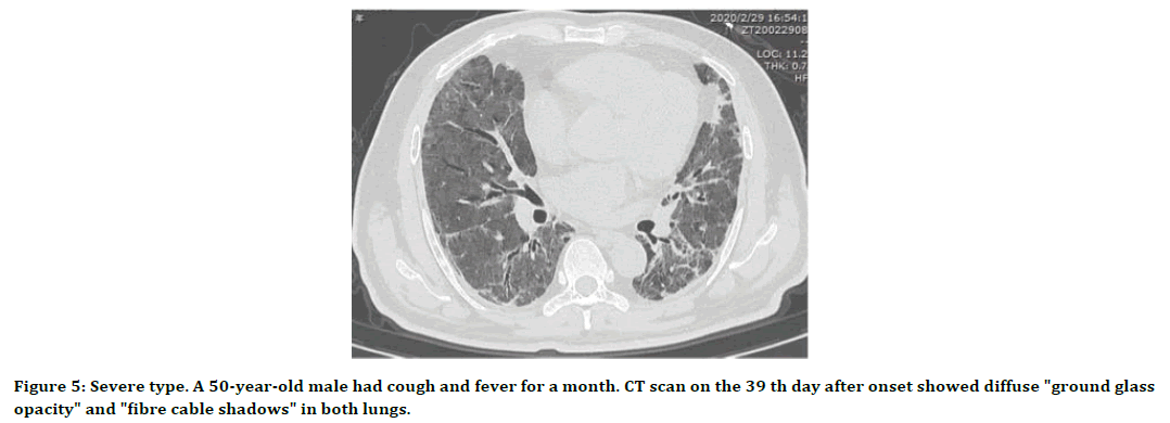
Figure 5. Severe type. A 50-year-old male had cough and fever for a month. CT scan on the 39 th day after onset showed diffuse "ground glass opacity" and "fibre cable shadows" in both lungs.
References
- WHO/Novelcoronavirus (China ) World Health Organization; Geneva : 2020.
- http://www.who.int /csr/don/05-Januar -2020-pneumonia -of-Unknown-cause-China/en
- Zhu N, Zhang D, Wang W, et al. A novel coronavirus from patients with pneumonia in China, 2019. New England J Med 2020.
- Amalou A, Türkbey B, Sanford T, et al. Targeted early chest CT in COVID-19 outbreaks as diagnostic tool for containment of the pandemic–A multinational opinion. Diagn Interv Radiol 2020; 26:292.
- Rodrigues JC, Hare SS, Edey A, et al. An update on COVID-19 for the radiologist-A British society of thoracic imaging statement. Clin Radiol 2020; 75:323-325.
- Rubin GD, Ryerson CJ, Haramati LB, et al. The role of chest imaging in patient management during the COVID-19 pandemic: A multinational consensus statement from the Fleischner society. Radiology 2020; 296:172-180.
Indexed at, Google Scholar, Cross Ref
Indexed at, Google Scholar, Cross Ref
Indexed at, Google Scholar, Cross Ref
Author Info
Ravi Bastakoti1, Adarsh Jha2*, Rabin Raj Singh1, Trinetra Kumar Karna1, Kshitij Giri1, Sunil Regmi1 and Atul Dwivedi3
1Koshi Hospital, Biratnagar, Nepal, India2Vedanta Hospital, Itahari, Nepal, India
3Shivaanan Polyclinic, Bareilly, Uttar Pradesh, India
Received: 11-Feb-2022, Manuscript No. JRMDS-22-53383; , Pre QC No. JRMDS-22-53383 (PQ); Editor assigned: 14-Feb-2022, Pre QC No. JRMDS-22-53383 (PQ); Reviewed: 28-Feb-2022, QC No. JRMDS-22-53383; Published: 07-Mar-2022
