Research - (2019) Volume 7, Issue 6
Clinical Case of Root Resorption Due to Improper Orthodontic Treatment
Artak G Heboyan1* and Anna A Avetisyan2
*Correspondence: Artak G Heboyan, Department of Prosthodontics, Yerevan State Medical University, Armenia, Email:
Abstract
The article presents a case of tooth root resorption due to improper orthodontic treatment. Before the orthodontic treatment, it is necessary to diagnose the anomaly precisely, to plan and correspondingly carry out a proper treatment, considering the direction, magnitude and duration of the orthodontic forces, individual peculiarities and risk factors in every clinical case to avoid complications and achieve a successful outcome. All above-mentioned, if not taken into consideration, leads to various complications such as root resorption. The peculiarity of the presented clinical case is that X-ray examination was neglected both at the stage of diagnosis and treatment planning and during treatment. The treatment lasted more than three years. As a result, the patient developed certain complaints and presented to our clinic with loose teeth and toothache. After appropriate examinations, root resorption was diagnosed in a number of teeth. Moreover, the root resorption was severely pronounced in maxillary central incisors which made it impossible to preserve the teeth. After the orthodontic treatment was completed, restoration of the teeth with implants was suggested. Root resorption conditioned by orthodontic treatment is diagnosed mostly at early stages and is possible to be prevented. However, this case is unique in its nature since multiple teeth are affected and the roots of some teeth are almost completely resorbed in one person. The objective of this paper is to emphasize the importance of orthodontic treatment planning, following conventional treatment protocols and proper application of orthodontic forces to prevent root resorption and achieve successful outcome.
Keywords
Root resorption, Orthodontic treatment, Orthodontic forcesIntroduction
The force, applied during orthodontic treatment, is a causative factor of inflammation and root resorption [1]. An optimal force of 20-150 g is needed to move the tooth and to reduce the severity of tooth resorption [2]. Excess force can cause periodontal ischemia in adults, since higher apical load occurs due to a thicker layer of cement as compared to that in children [3]. Compression on periodontal ligaments appearing during tooth movement increases the risk of root resorption which can be reduced and recover after the orthodontic forces are removed. Root resorption is inevitable in orthodontics, usually occurring less than 2.5 mm [4]. In pronounced root resorption, the tooth becomes loose, risking the whole process of treatment. The treatment methods can be modified to reduce root resorption during orthodontic treatment.
It is crucial to assess the risk factors of root resorption before treatment [5]. Numerous factors which contribute to the root resorption include the patient’s genetic predisposition and general health status. The occurrence of apical root resorption might be associated with single nucleotide variations in human genome, which assumes that orthodontic treatment is not the only factor in charge [6]. Thereby, risk factors are gender, age, genetic predisposition, some somatic diseases, hormonal disorders, mechanical trauma, tooth whitening, root abnormal shape, malocclusion, excessive orthodontic force, long-term orthodontic treatment [7].
Types of teeth differ in their susceptibility to resorption. Maxillary and lateral incisors are frequently subject to orthodontically induced apical resorption. Root resorption incidence occurring during orthodontic treatment ranges within 4-91% [8].
The article presents a case of root resorption due to improper orthodontic treatment. The objective of this paper is to emphasize the importance of orthodontic treatment planning, following conventional treatment protocols and correct application of orthodontic forces to prevent root resorption and achieve successful outcome.
Case Report
A 34-year-old female presented to the clinic with toothache and loose teeth. There was no concomitant disease mentioned in the patient’s medical history. The patient had been undergoing orthodontic treatment for three years. Extra oral examination revealed no facial asymmetry or any pathologic changes.
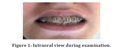
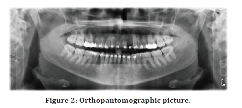
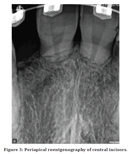
On intraoral examination, there were braces placed on the teeth (Figure 1). Intraoral examination revealed first degree tooth mobility and discoloration of the maxillary anterior teeth. Maxillary left lateral incisor was missing. Tooth percussion test was positive, no pulp response to the cold and hot stimuli was observed. Periodontal probing revealed the presence of pockets with 8 mm of depth in maxillary right canine and premolar. Orthopantomography showed that the patient was missing left lateral incisor, almost complete root resorption was observed in central incisors. Apical root resorption of the maxillary premolars, right canine and lateral incisor was visible on the orthopantomographic image. Roentgenography revealed interradicular bone resorption in maxillary right canine and premolar (Figure 2). Focused periapical roentgenography of central incisors was also carried out in order to avoid incorrect interpretation of the orthopantomographic images. These confirmed the presence of almost complete root resorption (Figure 3).

Figure 1: Intraoral view during examination.

Figure 2: Orthopantomographic picture.

Figure 3: Periapical roentgenography of central incisors.
Results
Taking into consideration the severity of root resorption in central incisors, the treatment recommended to the patient included their extraction after the orthodontic treatment, with further reconstruction of central incisors and missing lateral incisor by implants.
Discussion
The roots of maxillary central and lateral incisors are more liable to orthodontically induced apical resorption. In this case, the roots of central incisors were resorbed completely as compared to other teeth. Root resorption of lesser severity is observed in patients who underwent treatment at the age of 11 years and develops mostly in patients aged above 20 years [9]. It is more unlikely to occur in case tooth movement is completed before the full development of the roots. In the presented clinical case, the age of the patient appeared to be favorable factor for the occurrence of root resorption.
The magnitude of the force exerted and its intermittent application, causing less damage as compared to the continuous force applied, are considered to be the decisive factors in root resorption. Providing time for the reparative mechanism to act, intermittent force induces less root resorption. Root resorption is caused by the heavy force applied during the orthodontic treatment [10]. Long-term orthodontic treatment also contributes to root resorption. A month of extra treatment duration leads to 0.1-0.2 mm of additional root resorption. Average treatment duration in patients without root resorption is 1.5 years. The risk of severe root resorption increases if total treatment period exceeds 30 months. In the case, the patient had been undergoing the treatment for more than three years before attending our clinic.
Radiographic assessment of root resorption is common in clinical practice and is performed in the first 3–6 months and then every year after installation. In this case, no radiography was carried out either before or during treatment, being a crucial mistake. Eventually, resorption occurs at the active stage of orthodontic treatment. Temporary discontinuance of orthodontic treatment for 3 months is recommended at the first radiographic signs of resorption.
Root resorption treatment strategy depends on the resorption severity, being the most important factor. When resorption is diagnosed at the early stage, the application of orthodontic forces is temporarily discontinued. Pulp vitality is determined. Endodontic treatment is carried out if the pulp is infected.
There are various medicines to treat root resorption, e.g. Echistatin, which inhibits resorptive activity of isolated clastic cells. Local clodronate (Bisphosphonates), Prednisolone, Celecoxib suppress root resorption occurring during tooth movement. Root resorption induced by orthodontic force applied is considerably inhibited by different doses of risedronate administered locally. Calcium ions together with PGE2 stabilize root resorption. Nabumetone reduces external root resorption and pain caused by intrusive orthodontic movement.
Conclusion
In markedly pronounced resorption the tooth is extracted. No roentgenographic investigations were carried out to diagnose, plan the treatment and control the treatment process in this case. As a result, root resorption was diagnosed at late stage with no possibility of preserving central incisors. Thus, orthodontic treatment planning, proper application of orthodontic forces and following conventional treatment protocols allow avoiding root resorption and contributing to successful outcome.
References
- Abuara A. Biomechanical aspects of external root resorption in orthodontic therapy. Med Oral Patol Oral Cir Bucal 2007; 12:610-613.
- https://onlinelibrary.wiley.com/doi/book/10.1002/9781118916148
- Vikram NR, Kumar KSS, Nagachandran KS, et al. Apical stress distribution on maxillary central incisor during various orthodontic tooth movements by varying cemental and two different periodontal ligament thicknesses: A FEM study. Indian J Dent Res 2012; 23:213-220.
- https://link.springer.com/book/10.1007/978-3-319-18353-4
- Maues CPR, Nascimento RRD, Vilella ODV. Severe root resorption resulting from orthodontic treatment: Prevalence and risk factors. Dental Press J Orthod 2015; 20:52-58.
- Al-Qawasmi RA, Hartsfield JK, Evrett ET, et al. Genetic predisposition to external apical root resorption. Am J Orthod Dentofacial Orthop 2003; 123:242-252.
- Heboyan A, Avetisyan A, Markaryan M, et al. Tooth root resorption conditioned by orthodontic treatment. Oral Health Dental Sci 2019; 3;1-8.
- Motokawa M, Sasamoto T, Kaku M, et al. Association between root resorption incident to orthodontic treatment and treatment factors. Eur J Orthod 2012; 34:350-356.
- Motokawa M, Terao A, Kaku M, et al. Open bite as a risk factor for orthodontic root resorption. Eur J Orthod 2013; 35:790-795.
- Travess H. Orthodontics. Part 6: Risks in orthodontic treatment. Br Dent J 2004; 196:71-77.
Author Info
Artak G Heboyan1* and Anna A Avetisyan2
1Department of Prosthodontics, Yerevan State Medical University, Armenia2Department of Therapeutic Stomatology, Yerevan State Medical University, Armenia
Citation: Artak G Heboyan, Anna A Avetisyan, Clinical Case of Root Resorption Due to Improper Orthodontic Treatment, J Res Med Dent Sci, 2019, 7(6):91-93
Received: 30-Oct-2019 Accepted: 09-Dec-2019 Published: 16-Dec-2019
