Case Report - (2021) Volume 9, Issue 2
Bicornuate Unicollis Uterus with Gestation: A Rare Case Report
Prabu D, T. Ramachandra Prasad and Prabakaran M*
*Correspondence: Prabakaran M, Department of Radiodiagnosis, Sree Balaji Medical College and Hospital, Bharath Institute of Higher Education and Research, Chennai, India, Email:
Abstract
The study of disease transmission in general, innate uterine abnormalities happen in ~1.5% of females (run 0.1-3%). Bicornuate uteri are thought to speak to ~25% (territory 10-39%) of Mullerian conduit inconsistencies.
Keywords
Bicornuate unicollis uterus, Gestation, Bicornuate bicollis uterus, Ultrasound, USG pelvis
Introduction
A Bicornuate uterus is a sort of uterine duplication inconsistency. It tends to be delegated a class IV Mullerian conduit anomaly. A Bicornuate uterus is a typical kind of innate uterine inconsistency, and it conveys an expanded danger of barrenness and unnatural birth cycle [1-5].
Pathology
A bicornuate uterus results from a strange improvement of the paramesonephric channels. There is a halfway disappointment of combination of the pipes, bringing about an uterus isolated into two horns.
Subtypes
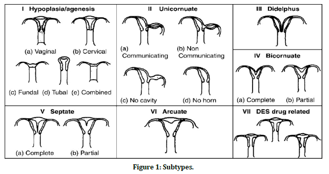
Bicornuate uterus is separated by the inclusion of the cervical channel (Figure 1):

Figure 1. Subtypes.
Bicornuate bicollis: two cervical waterways; focal myometrium stretches out to outside cervical os.
Bicornuate unicollis: one cervical waterway; focal myometrium stretches out to inner cervical os.
Radiographic features
General
The favored techniques for imaging uterine oddities are ultrasound, hysterosalpingogramor MRI. The outside uterine form is sunken or heart-molded, and the uterine horns are broadly disparate. The fundal split is ordinarily more than 1cm profound and the intercornual separation is extended. The uterus is viewed as involving caudally melded symmetric uterine holes with some level of correspondence between the two depressions (generally at the uterine isthmus). Despite the fact that not a particular finding, the edge between the horns of the bicornuate uterus is typically more than 105°3.
Hysterosalpingogram
HSG could not be done since patient was pregnant.
Ultrasound
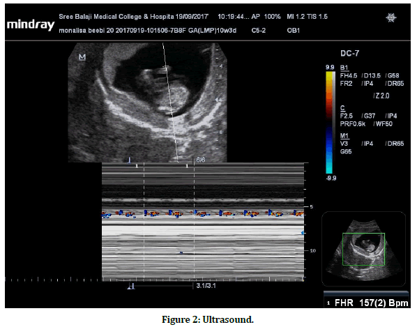
USG pelvis uncovered endometrium in the district of collection of uterus bifurcating cranially into two horns with interceding tissue reliable with typical myometrium. Single gestational sac with a fetal shaft of 12 weeks gestational age was available in the left horn and endometrial response was noted in the privilege horn. Fetal heart action was seen (Figure 2).

Figure 2. Ultrasound.
X-ray
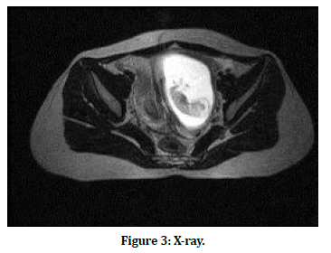
May help affirm life structures by demonstrating a profound (>1 cm) fundal separated in the external uterine shape and an intercornual separation of >4 cm. The uterus exhibits typical uterine zonal anatomy. MRI imaging uncovered a bicornuate uterus with gestation in the left horn and endometrial reaction in the right horn (Figure 3).

Figure 3. X-ray.
Treatment and prognosis
Careful intercession is typically not demonstrated without conceptive troubles. In ladies with a background marked by intermittent pregnancy misfortune and in whom no other barrenness issues have been recognized a Strassman metroplasty can be considered.
Differential analysis
Uterus didelphys: Complete disappointment of combination happens during the improvement of the paramesonephric conduits with duplication of the uterus, cervix and vagina Septate uterus 12: has an ordinary fundal shape however is portrayed by a constant longitudinal septum that in part partitions the uterine depression.
Discussion
Congenital uterine anomalies have been clearly implicated in women suffering with recurrent miscarriage. In women with infertility the role of these anomalies remains unclear. Correct assessment of prevalence of these anomalies in women with recurrent miscarriage and infertility will play a major role in the treatment. My case report is regarding a 20 yr old female having Bicornuate unicollis uterus with 12 weeks gestation. It was a natural conception after 2 years of married life. The ultrasound findings were normal for the gestational age. The gestation was in left horn of uterus with endometrial reaction in the right horn. I could not find any abnormalities in the ovaries.
Conclusion
Though Ultrasound is the primary investigation which is easily available, cheap and nonradiation to screen and detect uterine anomalies, Magnetic Resonance Imaging is more specific and sensitive for detection of Septate Uterus than Ultrasound.
Ethical Clearence
Nil.
Source of Funding
Self-funded.
Conflict of Interest
Nil.
References
- Graupera B, Pascual MA, Hereter L , et al. Accuracy of three-dimensional ultrasound compared with magnetic resonance imaging in diagnosis of mullerian duct anomalies using ESHRE-ESGE consensus on the classification of congenital anomalies of the female genital tract. Ultrasound Obstet Gynecol 2015; 46:616-622.
- Imboden S, Muller M, Raio L, et al. Clinical significance of 3D ultrasound compared to MRI in uterine malformations. Ultraschall Med 2014; 35:440-444.
- Saravelos SH, Cocksedge KA, Li TC. Prevalence and diagnosis of congenital uterine anomalies in women with reproductive failure: A critical appraisal. Hum Reprod Update 2008; 14:415-429.
- Bermejo C, Martinez Ten P, Cantarero R, et al. Three dimensional ultrasound in the diagnosis of mullerian duct anomalies and concordance with magnetic resonance imaging. Ultrasound Obstet Gynecol 2010; 35:593-601.
- Canzone G, Parlato M, Triolo L. 2D-3D Ultrasound in the diagnosis of uterine malformations. Donald School J Ultrasound Obstetr Gynecol 2007; 1:77-79.
Author Info
Prabu D, T. Ramachandra Prasad and Prabakaran M*
Department of Radiodiagnosis, Sree Balaji Medical College and Hospital, Bharath Institute of Higher Education and Research, Chennai, IndiaCitation: D Prabu, T.Ramachandra Prasad, M Prabakaran, Bicornuate Unicollis Uterus with Gestation–A Rare Case Report, J Res Med Dent Sci, 2021, 9 (2): 226-228.
Received: 09-Dec-2020 Accepted: 25-Jan-2021
