Research Article - (2022) Volume 10, Issue 5
Antimicrobial Activity of Ginger Silver Nano-composite on Cat-Gut Suture Material an In-Vitro Study
Guy Patrick Sandou1, Sahana3, Subhashree R2*, Thiyaneswaran Nesappan2 and S Rajeshkumar3
*Correspondence: Subhashree R, Department of Maxillofacial Prosthodontics and Oral Implantology, Saveetha Dental College and Research Institute, Chennai, India, Email:
Abstract
In this present study, silver nanoparticles were synthesized using dried ginger extract and intervened with chitosan to result in the silver nano-composite. The biosynthesized nano-composite was coated on catgut suture material to test its anti periimplantitis effect. The synthesized silver nanoparticles were characterized using UV-Visible spectrophotometry and showed peak absorption at 440 nm, and the composite showed at 380 nm. The TEM image clearly showed the uniformly dispersed spherical shaped silver nano-composite with the size of about 14-20 nm. The coated suture material was tested for its antiperi implantitis effect, and it showed a potent effect against S. mutans and E. feacalis.
Keywords
Dried ginger extract, Silver nanoparticles, Nano-composite, Catgut suture, Peri-implantitis
Introduction
Nanotechnology addresses a progressive way for innovative advancement that leads to material administration at the nanometer scale (one billion times less than a meter). Nanotechnology genuinely implies any innovation on the nanoscale that has various applications in reality. In a real sense, nanotechnology envelops the manufacture and utilisation of chemical, physical, and cellular systems at scales going from singular particles or molecules to submicron measurements and the coordination of these subsequent nanomaterials into more powerful frameworks. It can change our perspective and assumptions and give us the capacity to determine worldwide issues [1].
Nanoparticles are synthesised depending on various methodologies which incorporate chemical and biological methods. The silver nanoparticles are the most focused interest for research because of their potential in applications likes to change the physical, electronic and optical properties of a substance or compound, therapy of malignant growth, as clinical gadgets, antimicrobial coatings of paint, materials, additionally bio-sensing, likewise have mitigating, antiviral, antifungal and antiplatelet activities. The organic strategies are currently broadly being utilised in light of the fact because it is profoundly harmful and not environment friendly. The interest in utilising an eco-accommodating technique for nanoparticle synthesis has prompted another methodology, "green nanotechnology", prepared without using toxic chemicals [2].
Peri-implantitis is an inflammatory response with loss of supporting bone in the tissues encompassing an implant [3,4]. The general recurrence of peri-implantitis was accounted for to be 5% to 8% for chosen embed systems. An intersection of the gingival tissue with the neck of dental implants may forestall microorganisms’ colonisations prompting peri-implantitis. Dried ginger extract was used to analyse the better suture material for periimplantitis treatment in this study to synthesise silver nanocomposite [5].
Zingiber officinale (ginger) is a significant rhizome in Ayurvedic and Unani and Chinese medication, which is treated for different ailments like stomach-related illness, stiffness, and bleeding disorders because of a wide variety of unpredictable oils like zingiberol, monoterpene, sesquiterpene, and sesquiterpene hydrocarbons [6].
In this present study, dried ginger extract mediated silver nanocomposite was synthesised and coated on catgut suture material to test its anti-periimplantitis effect [7].
Materials And Methods
The chemical used in this study, such as silver nitrate (AgNO3), chitosan was from Sigma Aldrich, and microbial media, such as Mueller Hinton Agar, Rose Bengal Agar, were from Hi-Media Laboratories Pvt. Ltd. The Peri-implantitis causing pathogens such as Pseudomonas, S. mutans, E. faecalis, C. Albicans was isolated from patients at Saveetha Dental College and Hospitals, Poonamallee Chennai. The rhizome of Zingiber officinale (Ginger) was bought from a supermarket near Poonamallee. The ginger was washed thoroughly under tap water to clear away the remnants present on its surface [8]. It was shade dried for 7-8 days to attain dried form. Then the dried ginger was made into small pieces and grounded to a fine powder. The dried ginger powder was stored in an airtight container for further experiments [9].
Preparation of ginger extract0.5 g of dried ginger powder was measured and dissolved in 50 mL of distilled water and boiled for 10-15 minutes at 60-70ºC using a heating mantle. The boiled mixture was filtered using what man No: 1 filter paper. The filtered, dried ginger extract was used to prepare silver nanoparticles [10].
Synthesis of dried Ginger extract mediated silver nanoparticles1 mM of Silver nitrate (AgNO3) was dissolved in 90 mL of distilled water. To that, 10 mL of the filtered, dried ginger extract was added. The reaction mixture was kept in a magnetic stirrer at 600-700 rpm for 48 hours. The colour change was observed periodically, and the synthesis was confirmed using a UV-Visible double beam spectrophotometer [11]. The synthesized silver nanoparticles solutions were subjected to centrifugation at 8000 rpm for 10 minutes. The supernatant was discarded after the centrifuge, and the pellet was collected. The collected pellet was stored in an Eppendorff tube at 4ºC for characterization and other studies [12].
Synthesis of dried ginger extract mediated AgNPs based nanocomposite0.5 g of chitosan was measured and added to 49.5 mL of distilled water. To that, 0.5 mL of glacial acetic acid was added in order to prepare the nanocomposite. The chitosan mixture was stirred continuously for about 2-3 hours using a magnetic stirrer at 800 rpm to form a clear solution. 50 mL of pre-centrifuged dried ginger extract AgNPs solution was added to 50 mL of chitosan solution. This nanocomposite solution was stirred using a magnetic stirrer at 800 rpm for three days [13].
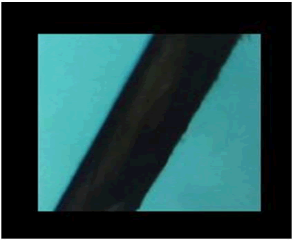
Coating dried ginger extract mediated AgNPs based nano-composite on cat-gut suture materialTo the biosynthesized nano-composite solution, sterile catgut suture material was placed, and the coating process was done by stirring at 800-950 rpm and heating at 50ºC using a magnetic stirrer. The catgut suture material along with biosynthesized AgNPs based nanocomposite solution was further dried and coated by placing it in a sterile glass Petri plate and kept inside a hot air oven at 60-70ºC for 6 hours. The dried and completely coated suture material was observed under the digital microscope to check its coated surface with control catgut suture material. The coated catgut suture material was stored in a sterile Eppendorff tube for further experiments (Figure 1).

Figure 1: coated catgut suture.
Characterization studiesThe size and shape of the green synthesized silver nanoparticles were analyzed by using Transmission Electron Microscopy (TEM), and the synthesis of silver nanoparticles was confirmed by using a UV-Visible double beam spectrophotometer in the wavelength range of 250-750 nm.
Biomedical Application
Antimicrobial activityThe antimicrobial activity was done using the agar well diffusion technique. 10 µL of fresh bacterial cultures such as Pseudomonas, S. mutans, E. faecalis and C. albicans were inoculated in a sterile Hi-veg broth medium and incubated for 18 hours in an orbital shaker at 120-150 rpm. Mueller Hinton agar was prepared. The antimicrobial activity was done to analyse the efficacy of dried ginger extract mediated AgNPs based nanocomposite on cat-gut suture material. The pathogens are swabbed on the surface of the agar plates using a sterile cotton swab. The coated catgut suture material is placed on the surface of the swabbed MHA plates along with a control group for each respective plate [14].
The plates were incubated at 37°C for 24 hours (except for C. Albicans, the plates were incubated at 37°C for 48 hours), and the zone of inhibition is measured around the suture material to compare and study the potential anti-periimplantitis activity [15].
Result
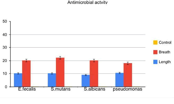
Preventing peri- implantitis is an important parameter for obtaining a desired outcome. The study results demonstrated that the catgut suture coated with ginger silver nanoparticles material compared with the control group, held a potent antimicrobial effect against all four pathogens, which increased its effect in a dose-dependent manner, and it was depicted. A significant zone of inhibition in the agar well culture method was found in E. fecalis and S. mutans than other two organism’s C. albicans and Pseudomonas. The zone of inhibition showed highest in S. mutans and Pseudomonas the least with respect to breath. Zone of inhibition showed least in S. albicans when compared to other organisms (Figure 2).

Figure 2: Antimicrobial activity nano-composite coated material.
Visual observationVisual observation was used as an initial identification tool to confirm the synthesis of nanoparticles. The initial colour of the biosynthesized silver nanoparticles solution was pale yellow, and it gradually changed to dark brown in colour after 2 hours. This colour change indicates the reducing ability of dried ginger extract, which leads to the reduction from silver nitrate to silver nanoparticles.
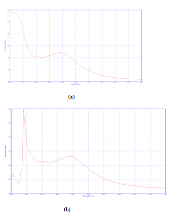
UV-Visible spectrophotometryUV-Visible spectrophotometry was used to detect the synthesis of nanoparticles at various intervals of time. Figure 3 (a) represents the absorption spectra of a biosynthesized silver nanoparticle which obtained its sharp absorption peak at 385 nm. The absorption peak at 380 nm is due to the excitation of surface Plasmon resonance which further confirms reduced silver ions by the dried ginger extract. Existing works are also reported to obtain the absorption peak at 440 nm for silver nanoparticles synthesized using Aloe vera and neem.
The UV-Visible spectra of dried ginger mediated AgNPS based nanocomposite were depicted in figure 3 (b) in the wavelength range of 250-750 nm. The biosynthesized Ag nanocomposite showed synergistic bands at around 440 nm for silver nanoparticles, blue-shifted to 380 nm in the composite. The UV results of the biosynthesized silver nanocomposite correlate with works that deal with Ag-MgO nanocomposite synthesis by using aqueous peel extract of Citrus paradise (Figure 3).

Figure 3: (a) UV-Visible spectra of dried ginger extract mediated silver nanoparticles
(b) UV-Visible spectra of dried ginger extract mediated AgNPs based nanocomposite.
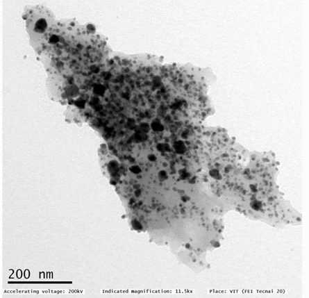
TEMTransmission Electron Microscope was assisted in analysing the shape and size of the synthesized silver nanocomposite, depicted. The nanocomposite shape was found to be spherical and uniformly dispersed in nature and size of about 14-20 nm. The TEM image also showed that the silver nanoparticles intercalated on the chitosan surface. Recently, researchers synthesized graphene oxide silver nanocomposite and the size of the nanocomposite lies around 18 nm (Figure 4).

Figure 4: TEM image of green synthesized Ag nanocomposite.
Antimicrobial activityThe antimicrobial effect of biosynthesized silver nanocomposite coated catgut suture material was tested against four peri-implantitis causing pathogens such as Pseudomonas, S. mutans, E. faecalis, C. albicans. A maximum zone of inhibition was observed in S. mutans and E. faecalis, among the other two organisms. This silver nanocomposite represents an attractive alternative for treating peri-implantitis in the dental field rather than using commercial antibiotics. Existing works closely related to silver micro fibrillated cellulose biocomposite once again confirms the intercalation of chitosan and silver nanoparticles that seems to hold an excellent antimicrobial effect.
Discussion
Preventing any post-surgical infection is an essential parameter for obtaining the desired outcome. This study showed that ginger Ag coated sutures showed an effective antimicrobial action against L. bacilli, S. mutans and C. albicans, which are the most commonly found oral microbiota.
Antimicrobial herbal traditional therapies or plants range from the rapidly growing flora of woods to those grown in farm fields. They have been studied, examined, proven to have antimicrobial properties, or are being checked now. All around the world, these herbs grow spontaneously or are cultivated primarily for this purpose. As a result of advances in herbal medicine, new and popular herbs have arisen. Incorporating herbal extracts into nanomedicine has several additional benefits, including bulk dosing and the ability to counteract absorption, which is a significant issue, attracting the interest of major pharmaceutical companies.
If left untreated, pathogens can cause a wide range of human diseases, from self-limiting illnesses to potentially lethal medical conditions. Nonetheless, due to widespread use, antibiotic resistance is on the rise, leaving previously treatable infections untreatable. Globalisation also facilitated the proliferation of (resistant) strains of other infectious agents. Recent research in the field of newer antibiotic therapies is best suitable for antibiotic resistance. In our study, ginger and mint silver nanoparticle antimicrobial activity was analysed using the agar well diffusion method and outcomes were analysed by a zone of inhibition.
UV-Visible spectrophotometer was used to detect the synthesis of nanoparticles at various intervals of time. Existing works are also reported to obtain the absorption peak at 440 nm for silver nanoparticles synthesized using Aloe vera and neem. The UV results of the biosynthesized silver nanocomposite correlate with works that deal with Ag-MgO nanocomposite synthesis by using aqueous peel extract of citrus paradisi. Recently, researchers synthesized graphene oxide-silver nanocomposite and the size of the nanocomposite lies around 18nm.
Studied the antibacterial properties of ginger nanoparticles. It was tested against Staphylococcus aureus, Klebsiella pneumonia, Candida albicans, Bacillus subtilis, Erwinia carotovora, and Proteus Vulgaris, as well as antifungal properties against Candida albicans. Antimicrobial analyses show that these nanoparticles have solid antimicrobial activity compared to antibiotic norm performance. experimented with silver nanoparticles using ginger extract against aquatic microorganisms, and results showed significant antimicrobial effects against typical aquatic pathogenic strains of Vibrio anguillarum, Vibrio alginolyticus, Aeromonas punctata, Vibrio parahaemolyticus, Vibrio splendidus and Vibrio harveyi.
Preventing peri-implantitis is an important parameter for obtaining a desired outcome. The antimicrobial effect of biosynthesized silver nanocomposite coated catgut suture material was tested against four peri-implantitis causing pathogens such as Pseudomonas, S. mutans, E. faecalis, C. albicans. A maximum zone of inhibition was observed in S. mutans and E. faecalis, among the other two organisms. This silver nanocomposite represents an attractive alternative for treating peri-implantitis in the dental field rather than using commercial antibiotics. Existing works closely related to silver micro fibrillated cellulose biocomposite once again confirms the intercalation of chitosan and silver nanoparticles that seems to hold an excellent antimicrobial effect.
While nanopharmaceuticals may appear to provide limitless possibilities in the field of drug delivery for the diagnosis and treatment of a variety of diseases, their safety should not be overlooked. Engineered nano-sized materials' physicochemical and structural properties can change as their size decreases, resulting in various material interactions that could lead to toxicological effects. Scientists must agree that the toxicological assessment of nanomaterials and nanomedicines is still in its early stages, and there are few reports on their safety and toxicity.
Conclusion
Catgut suture material has high tensile strength and can be easily absorbed by our body. Due to its unique features, it has been used in many surgical procedures. To make the suture material more resistant against peri-implantitis causing pathogens, dried ginger extract mediated silver nano-composite has been developed. The nano-composite showed a uniformly dispersed spherical shape and with a size of about 14-20 nm in the TEM image. The developed suture material showed an excellent antimicrobial effect on all tested pathogens, revealing its significant role in future biomedical applications in the dental field.
References
- Rajeshkumar S, Tharani M, Jeevitha M, et al. Anticariogenic Activity of Fresh Aloe Vera Gel Mediated Copper Oxide Nanoparticles. Indian J Public Health Res Dev 2019; 10.
- Arsath NM, Karunagaran M, Rajeshkumar S. Green synthesis and characterization of silver nanoparticle using phyllanthus emblica and cinnamomum verum extract. Plant Cell Biotechnol Mol Biol 2020;120-6.
- Menon S, Agarwal H, Kumar SR, et al. Green synthesis of silver nanoparticles using medicinal plant Acalypha indica leaf extracts and its application as an antioxidant and antimicrobial agent against foodborne pathogens. Int J Appl Pharm 2017; 9:42-50.
- Berglundh T, Persson L, Klinge B. A systematic review of the incidence of biological and technical complications in implant dentistry reported in prospective longitudinal studies of at least 5 years. J Clin Periodontol 2002; 29:197-212.
[Crossref] [Google Scholar] [Pubmed]
- Banerjee S, Mullick HI, Banerjee J, et al. Zingiber officinale: ‘a natural gold’. Int J Pharmaceutical Bio-Sci 2011; 2:283-294.
- Behera S, Ojha A, Rout J, Nayak P. Plant mediated synthesis of silver nanoparticles: opportunity and challenge. Int. j. biol. pharm. allied sci 2012; 1:1637-1658.
- Singh C, Sharma V, Naik PK, et al. A green biogenic approach for synthesis of gold and silver nanoparticles using Zingiber officinale. Dig J Nanomater Biostructures 2011; 6:535-542.
- Rajeshkumar S. Synthesis of silver nanoparticles using fresh bark of Pongamia pinnata and characterization of its antibacterial activity against gram positive and gram negative pathogens. Resource-Efficient Technologies 2016; 2:30-35.
- Asha S, Thirunavukkarasu P, Rajeshkumar S. Green synthesis of silver nanoparticles using mirabilis jalapa aqueous extract and their antibacterial activity against respective microorganisms. Res J Pharm Technol. 2017; 10:811-7.
- Chithralekha B, Rajeshkumar S. Cytotoxic effect of Aloe vera and neem herbal formulations assisted silver nanoparticles. Drug Invent Today 2019; 12.
- Ayinde WB, Gitari MW, Muchindu M, et al. Biosynthesis of ultrasonically modified Ag-MgO nanocomposite and its potential for antimicrobial activity. J Nanotechnol 2018.
- Kumari S, Sharma P, Yadav S, et al. A novel synthesis of the graphene oxide-silver (GO-Ag) nanocomposite for unique physiochemical applications. ACS omega 2020; 5:5041-5047.
[Crossref] [Google Scholar] [Pubmed]
- Garza-Cervantes JA, Mendiola-Garza G, de Melo EM, et al. Antimicrobial activity of a silver-microfibrillated cellulose biocomposite against susceptible and resistant bacteria. Sci Rep 2020; 10:1-7.
[Crossref] [Google Scholar] [Pubmed]
- Rajeshkumar S, Sherif MH, Malarkodi C, et al. Cytotoxicity behaviour of response surface model optimized gold nanoparticles by utilizing fucoidan extracted from padina tetrastromatica. J Mol Struct 2021; 1228:129440.
- Chellapa LR, Rajeshkumar S, Arumugham MIet al. Biogenic Nanoselenium Synthesis and Evaluation of its antimicrobial, Antioxidant Activity and Toxicity. Bioinspir Biomim Nan 2020:1-6.
Author Info
Guy Patrick Sandou1, Sahana3, Subhashree R2*, Thiyaneswaran Nesappan2 and S Rajeshkumar3
1Department of Implantology, Saveetha Dental College and Research Institute, Chennai, India2Department of Pharmacology, Saveetha Dental College and Research Institute, Chennai, India
3Department of Maxillofacial Prosthodontics and Oral Implantology, Saveetha Dental College and Research Institute, Chennai, India
Citation: Guy Patrick Sandou, Subhashree R, S Rajeshkumar, Thiyaneswaran Nesappan, Sahana, Antimicrobial Activity of Ginger Silver Nano-composite on Cat-Gut Suture Material an In-Vitro Study , J Res Med Dent Sci, 2022, 10(5): 118-122.
Received: 21-Feb-2022, Manuscript No. 48561; , Pre QC No. 48561; Editor assigned: 23-Feb-2022, Pre QC No. 48561; Reviewed: 09-Mar-2022, QC No. 48561; Revised: 22-Apr-2022, Manuscript No. 48561; Published: 06-May-2022
