Research Article - (2022) Volume 10, Issue 8
Anterior Tooth Fracture Among Patients Visiting Private Dental College: A Retrospective Study
Chris Noel Timothy, Arthi Balasubramaniam* and Sudharrshiny S
*Correspondence: Arthi Balasubramaniam, Department of Public Health Dentistry, Saveetha Dental College and Hospitals, Saveetha Institute of Medical and Technical Sciences (SIMATS), Saveetha University, Chennai, India, Email:
Abstract
Background: Traumatic dental injuries to the anterior teeth are common occurrences and affect approximately 20-30% of the permanent dentition all over the world. This often affects the aesthetics, phonetics and function. Although commonly occurring in the primary dentition, it also occurs in permanent teeth. Thus, this study aims to assess the prevalence of anterior teeth fracture among patients attending a private dental college.
Materials and Methods: This was a retrospective study, where all the data of the patients were obtained from the patient reports who visited a private dental college in Chennai, India. Data regarding the permanent anterior teeth fracture of the patients with age ranging from 10 to 70 years were collected and analyzed using IBM SPSS version 23.0.
Results: The prevalence of anterior teeth fracture was higher in males (68.9%) than in females (31.1%). Also, the prevalence was found to be high among the age group 21 to 40 years (24.4%). In this study we observed that there is no significant association of age and gender with type of tooth fracture in both the upper and lower anteriors (p>0.05).
Conclusion: Males were found to be more prone to anterior teeth fracture compared to females. The age group of 21 to 40 years were more commonly seen with anterior teeth fractures. Fractures were more commonly seen in the upper anteriors compared to the lower anteriors. Ellis class 1 or enamel fracture was found to be more prevalent.
Keywords
Anterior teeth, Ellis fracture, Restoration, Green energy green synthesis
Introduction
Dental trauma is an irreversible pathology which often has implications that extend beyond dentistry. Anterior teeth are most commonly affected. This occurs due to physical impact on the tooth which can either be direct or indirect. Regardless, the damage is usually permanent unless quick dental treatment is administered. A system of classification has been set in place for evident tooth fractures in the form of Ellis and Davey classification [1]. It includes class 1 which is a simple fracture of the crown involving only enamel, class 2 which is the extensive fracture of the crown involving both enamel and dentin, class 3 which is the extensive fracture of the crown involving enamel, dentin and pulp, class 4 where the tooth has become non-vital with or without the loss of crown structure, class 5 which is the complete loss of teeth as a result of trauma, class 6 which is the fracture of the root with or without the loss of crown structure, class 7 which is the displacement of the tooth without fracture of crown or root structure and finally class 8 which is the displacement of the crown en masse and its displacement [2-8].
Anterior dental trauma affects the function, aesthetics, speech and stability of the oral cavity. Dental malocclusion is a predisposing factor for tooth fracture, especially patients suffering from class 2 dental malocclusion [9]. Anterior teeth fracture not only affects patients physically but also psychologically where they become aware about their own looks which leads to a buildup in stress and anxiety [10-12]. A previous research by Rai, et al. it was found that among 4500 children, the most commonly affected teeth were the maxillary incisors, the etiology of which was undefined falls [13]. It was also found in another study conducted by Hegde et al, that greater the over jet, greater was the risk of traumatic injury to the anterior teeth. Patients with proclaimed maxillary incisors and canines possessed a 3.5-fold increase in risk of dental trauma [14,15].
Anterior tooth fracture was also more prevalent in urban population compared to the rural population; this could be due to occupational hazards, road traffic accidents, sporting injuries and violence [14]. Another reason why this research study is of importance is due to the fact that the majority of the substantial research studies surrounding anterior teeth fracture have majorly focused on the pediatric dental population and have ignored the adult population.
Thus, this research study is needed to assess the incidence and prevalence of anterior teeth fracture among the Chennai population. This study will fulfill the deficiency of a lack of prevalence data for permanent anterior teeth fracture. Our team has extensive knowledge and research experience that has translated into high quality publications [16-35]. The aim of the current study is to assess the prevalence of anterior teeth fracture among patients attending a private dental college.
Materials and Methods
Study setting and design
This research study was defined as a retrospective study where the entire patient’s data that reported to Saveetha Dental College and hospitals, SIMATS, Chennai, India and were diagnosed with anterior teeth fracture were obtained from the case records in the Dental Information Archiving Software (DIAS). This study setting was a university setting and the research study was conducted in the dental clinics of Saveetha Dental College. This setting came with various pros and cons. The pros included the presence of a larger population and an abundant availability of data. Some of the cons included the study taking place in an unicentric setting and possessing a very limited demographic.
Sample selection and criteria
A non-probability convenience sampling method was used to select the patient reports. This population was selected from the patients who visited the undergraduate and postgraduate dental clinics in Saveetha dental college. A total of 88,950 patient records were reviewed and cross verification was performed by an additional reviewer out of which 90 cases were found to have anterior teeth fracture. The age group of the patients ranged from 10-70 years. No gender restriction was placed. The minimization of sample bias was performed by an additional reviewer, acquiring all the data from within the university and as an additional measure. Sample collection was performed from March 2020 to March 2021. Any incomplete, duplicated or censored data that was present in the collected data was excluded from the study. The inclusion criteria for the current study included outpatients who underwent diagnosis and were found to have anterior teeth fracture whereas the exclusion criteria comprised patients who did not have anterior tooth fracture.
Ethical approval
The approval to undertake this research study had been approved by the ethical board of Saveetha University (applied).
Statistical analysis
The data was then arranged in a methodical manner using Microsoft excel software and was tabulated on the basis of 4 parameters namely, age of subject, gender of subject, fractures in the upper anterior and fractures in the lower anterior. Statistical analysis of the compiled data was performed using IBM SPSS statistical analyzer V.23.0. Descriptive statistics and Chi square test was done for statistical analysis. A statistical significance p value<0.05 was considered The dependent variables of the study included the prevalence of different classes of anterior teeth fracture. The Independent variables included the age and gender of the subjects.
Results
The data was collected and sorted based on the 4 parameters mentioned previously and Figure 1 explains about the age wise distribution of the study population. Most common age groups were 21 to 30 years and 31 to 40 years with 24.4% each, followed by 41 to 50 years with 18.9%, 10 to 20 years with 16.7%, 51 to 60 years with 12.2% and finally 61 to 70 years with 3.3%.
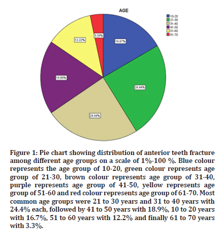
Figure 1: Pie chart showing distribution of anterior teeth fracture among different age groups on a scale of 1%-100 %. Blue colour represents the age group of 10-20, green colour represents age group of 21-30, brown colour represents age group of 31-40, purple represents age group of 41-50, yellow represents age group of 51-60 and red colour represents age group of 61-70. Most common age groups were 21 to 30 years and 31 to 40 years with 24.4% each, followed by 41 to 50 years with 18.9%, 10 to 20 years with 16.7%, 51 to 60 years with 12.2% and finally 61 to 70 years with 3.3%.
Figure 2 demonstrated the gender wise distribution of the study population. We observed that out of 90 cases, more male predilection was seen with 68.9% and females with 31.1%.
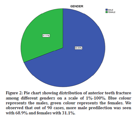
Figure 2: Pie chart showing distribution of anterior teeth fracture among different genders on a scale of 1%-100%. Blue colour represents the males, green colour represents the females. We observed that out of 90 cases, more male predilection was seen with 68.9% and females with 31.1%.
Figure 3 explains about the type of tooth fracture in relation to the upper anterior which include the teeth 11, 12, 13, 21, 22 and 23. In this we observed that the majority of the teeth presented with Ellis class 1 with 48.9% followed by Ellis class 2 with 42.2%, normal teeth with 4.4% and Ellis class 3 and 4 each with 2.2%.
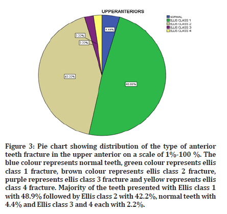
Figure 3: Pie chart showing distribution of the type of anterior teeth fracture in the upper anterior on a scale of 1%-100 %. The blue colour represents normal teeth, green colour represents ellis class 1 fracture, brown colour represents ellis class 2 fracture, purple represents ellis class 3 fracture and yellow represents ellis class 4 fracture. Majority of the teeth presented with Ellis class 1 with 48.9% followed by Ellis class 2 with 42.2%, normal teeth with 4.4% and Ellis class 3 and 4 each with 2.2%.
Figure 4 explains about the type of tooth fracture in relation to the lower anterior which include the teeth 31, 32, 33, 41, 42 and 43. In this we observed that the majority of the teeth presented with normal tooth form with 92.2% followed by Ellis class 1 with 5.6% and Ellis class 2 with 2.2%.
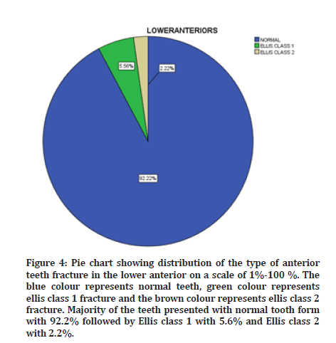
Figure 4: Pie chart showing distribution of the type of anterior teeth fracture in the lower anterior on a scale of 1%-100 %. The blue colour represents normal teeth, green colour represents ellis class 1 fracture and the brown colour represents ellis class 2 fracture. Majority of the teeth presented with normal tooth form with 92.2% followed by Ellis class 1 with 5.6% and Ellis class 2 with 2.2%.
Figure 5 explains the association between the age of the participants and the type of tooth fracture in the upper anterior. It was found that Ellis class 1 fracture was more frequent in the age groups of 21 to 30 years, 41 to 50 years and 51 to 60 years, whereas Ellis class 2 fractures were more commonly observed in the age groups of 10 to 30 years, 31 to 40 years and 61 to 70 years.
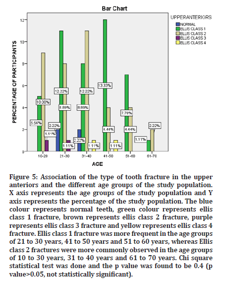
Figure 5: Association of the type of tooth fracture in the upper anteriors and the different age groups of the study population. X axis represents the age groups of the study population and Y axis represents the percentage of the study population. The blue colour represents normal teeth, green colour represents ellis class 1 fracture, brown represents ellis class 2 fracture, purple represents ellis class 3 fracture and yellow represents ellis class 4 fracture. Ellis class 1 fracture was more frequent in the age groups of 21 to 30 years, 41 to 50 years and 51 to 60 years, whereas Ellis class 2 fractures were more commonly observed in the age groups of 10 to 30 years, 31 to 40 years and 61 to 70 years. Chi square statistical test was done and the p value was found to be 0.4 (p value>0.05, not statistically significant).
Figure 6 explains the association between the age of the participants and the type of tooth fracture in the lower anterior. It was found that Ellis class 1 fracture was more commonly observed in the age group of 21 to 30 years.
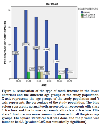
Figure 6: Association of the type of tooth fracture in the lower anteriors and the different age groups of the study population. X axis represents the age groups of the study population and Y axis represents the percentage of the study population. The blue colour represents normal teeth, green colour represents ellis class 1 fracture and the brown represents ellis class 2 fracture. Ellis class 1 fracture was more commonly observed in all the given age groups. Chi square statistical test was done and the p value was found to be 0.3 (p value>0.05, not statistically significant).
Figure 7 demonstrates the association between the gender of the participants and the type of tooth fracture in the upper anterior. It was found that Ellis class 1 and class 2 were more prevalent in males compared to females. Ellis class 3 and class 4 presented with similar prevalence in both males and females.
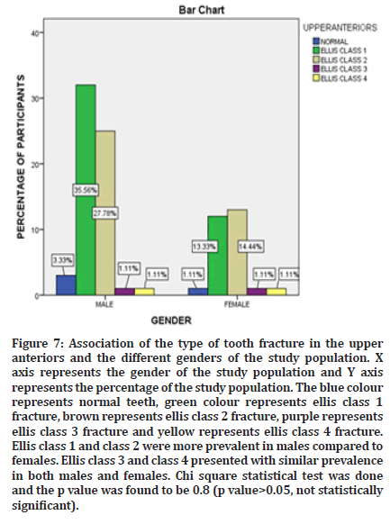
Figure 7: Association of the type of tooth fracture in the upper anteriors and the different genders of the study population. X axis represents the gender of the study population and Y axis represents the percentage of the study population. The blue colour represents normal teeth, green colour represents ellis class 1 fracture, brown represents ellis class 2 fracture, purple represents ellis class 3 fracture and yellow represents ellis class 4 fracture. Ellis class 1 and class 2 were more prevalent in males compared to females. Ellis class 3 and class 4 presented with similar prevalence in both males and females. Chi square statistical test was done and the p value was found to be 0.8 (p value>0.05, not statistically significant).
Figure 8 demonstrates the association between the gender of the participants and the type of tooth fracture in the lower anterior. It was found that Ellis class 1 fractures were more common in males compared to females.
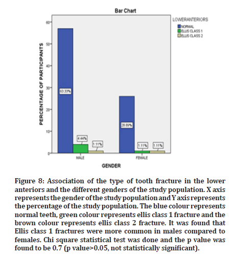
Figure 8:Association of the type of tooth fracture in the lower anteriors and the different genders of the study population. X axis represents the gender of the study population and Y axis represents the percentage of the study population. The blue colour represents normal teeth, green colour represents ellis class 1 fracture and the brown colour represents ellis class 2 fracture. It was found that Ellis class 1 fractures were more common in males compared to females. Chi square statistical test was done and the p value was found to be 0.7 (p value>0.05, not statistically significant).
Discussion
On review of literature, it was found that in a study conducted by Gopinath, et al. the most common ages where anterior tooth fracture was prevalent were 12 and 16 years which is not in concordance with the current study [36]. The reason for the male predominance could be due to a greater degree of occupational hazards, accidents with heavy machinery, sporting injuries, etc. but further research is required in order to prove this. In a recent study conducted by Abu-Hussein et al, it was found that males had experienced a significantly higher prevalence of trauma (8%) compared to females (4.2%) which is in concordance with our study [37]. The results of the current study also suggest that the prevalence of tooth fracture was greater in the maxillary anterior compared to the mandibular anterior which is in concordance with literature [38].
Previous research done similar to the current study includes a prevalence study performed by Olusegun, et al. where the prevalence of anterior teeth fracture was 0.4% in a study population of 1039 children. It was also found that incisors were the most commonly affected [39]. In another study performed by Mohan et al in Tamilnadu, it was found that, among 3200 children of 3 to 13 years of age, the prevalence rate was 10.13% and that the enamel fracture was the most commonly observed [40]. In a study conducted by Tasneem, et al. in a Kashmiri population, it was found that the rate of traumatic dental injuries in a study population of 1600 children was 9.3% [41]. In the current study the prevalence of anterior teeth fracture was found to be 0.001%.
Study Limitations
Presence of a smaller sample size along with the study being an unicentric one with a limited demographic and a lack of variety in the collected data.
Future Scope
This study could pave way for newer research with improved assessment and prevention of anterior dental fractures which will lead to better prognosis for the entire community.
Conclusion
There is no significant association between the anterior teeth fracture and the different age groups and gender. Males were found to be more prone to anterior teeth fracture compared to females. The age group of 21 to 40 years was more commonly seen with anterior teeth fractures. Fractures were more commonly seen in the upper anterior compared to the lower anterior. Ellis class 1 or enamel fractures were found to be more prevalent.
Acknowledgement
The authors would like to acknowledge the help and support rendered by the Saveetha dental college and hospitals for their constant assistance with the research and for granting us permission to access and utilize patient records for the purpose of this study.
Conflict of Interest
Authors declare no potential conflict of interest for this study.
Funding Support
The present project is sponsored by Saveetha Institute of Medical and Technical Sciences, Saveetha Dental College and Hospitals, Saveetha University.
References
- Ellis SG. Incomplete tooth fracture–proposal for a new definition. Br Dent J 2001; 190:424-428.
- Delattre JP, Resmond-Richard F, Allanche C et al. Dental injuries among schoolchildren aged from 6 to 15, in Rennes (France). Dent Traumatol 1995; 11:186-188.
- Kole D, Dholey MK, Sen S. Prevalence of permanent anterior teeth fracture among young children’s aged 8-14 years according to ellis and davey’s classification-An epidemiological study. J Dent Med Sci 2019; 18:24-29.
- Ahlawat B, Kaur A, Thakur G, et al. Anterior tooth trauma: A most neglected oral health aspect in adolescents. Indian J Oral Sci 2013; 4:31.
- Sharva V, Reddy V, Bhambal A, et al. Traumatic dental injuries to the anterior teeth among 12-year and 15-year-old schoolchildren of urban and rural areas of Bhopal District, Central India: A prevalence study. Chrismed J Health Res 2017; 4:38.
- Tiwari V, Saxena V, Tiwari U, et al. Dental trauma and mouthguard awareness and use among contact and noncontact athletes in central India. J Oral Sci 2014; 56:239-243.
- Mahajan N, Bansal S, Singla J. Glass fiber-reinforced composite post and core used for restoration of traumatically fractured anterior teeth. Indian J Oral Sci 2015; 6:73.
- Ellis RG, Davey KW. The classification and treatment of injuries to the teeth of children: a reference manual for the dental student and the general practitioner. Year B Med Publ 1970.
- Dosdogru EY, Görken FN, Erdem AP, et al. Maxillary incisor trauma in patients with class II division 1 dental malocclusion: Associated factors. J Istanb Univ Fac Dent 2017; 51:34-41.
- Arhakis A, Athanasiadou E, Vlachou C. Social and psychological aspects of dental trauma, behavior management of young patients who have suffered dental trauma. Open Dent J 2017; 11:41.
- Lee JY, Divaris K. Hidden consequences of dental trauma: the social and psychological effects. Pediatr Dent 2009; 31:96-101.
- Rao LN, Shetty A, Hedge MN. Psychological Effects of Trauma to Anterior Teeth. Exec Ed 2020; 11:125.
- Rai SB, Munshi AK. Traumatic injuries to the anterior teeth among South Kanara school children--a prevalence study. J Indian Soc Pedod Prev Dent 1998; 16:44-51.
- Hegde MN, Sajnani AR. Prevalence of permanent anterior tooth fracture due to trauma in South Indian population. Eur J Gen Dent 2015; 4:87-91.
- Hegde MN, Abootty S, Attavar S. Prevalence of anterior tooth fracture due to trauma. World J Dent 2015; 6:77-81.
- Mathew MG, Samuel SR, Soni AJ, et al. Evaluation of adhesion of Streptococcus mutans, plaque accumulation on zirconia and stainless steel crowns, and surrounding gingival inflammation in primary molars: Randomized controlled trial. Clin Oral Investig 2020; 24:3275-3280.
- Samuel SR. Can 5-year-olds sensibly self-report the impact of developmental enamel defects on their quality of life?. Int J Paediatr Dent 2020; 31:285-286.
- Samuel SR, Kuduruthullah S, Al Shayeb M. Impact of pain, psychological-distress, SARS-CoV2 fear on adults’ OHRQOL during COVID-19 pandemic. Saudi J Biol Sci 2021; 28:492-494.
- Samuel SR, Kuduruthullah S, Khair AMB, et al. Dental pain, parental SARS-CoV-2 fear and distress on quality of life of 2 to 6 year-old children during COVID-19. Int J Paediatr Dent 2021; 31:436-441.
- Samuel SR, Acharya S, Rao JC. School Interventions–based prevention of early-childhood caries among 3–5-year-old children from very low socioeconomic status: Two-year randomized trial. J Public Health Dent 2020; 80:51-60.
- Vikneshan M, Saravanakumar R, Mangaiyarkarasi R, et al. Algal biomass as a source for novel oral nano-antimicrobial agent. Saudi J Biol Sci 2020; 27:3753-3758.
- Chellapa LR, Shanmugam R, Indiran MA, Samuel SR. Biogenic nanoselenium synthesis, its antimicrobial, antioxidant activity and toxicity. Bioinspired Biomim Nanobiomaterial 2020; 9:184-189.
- Samuel SR, Mathew MG, Suresh SG, et al. Pediatric dental emergency management and parental treatment preferences during COVID-19 pandemic as compared to 2019. Saudi J Biol Sci 2021; 28:2591-2597.
- Barma MD, Muthupandiyan I, Samuel SR, et al. Inhibition of Streptococcus mutans, antioxidant property and cytotoxicity of novel nano-zinc oxide varnish. Arch Oral Biol 2021; 126:105132.
- Muthukrishnan L. Nanotechnology for cleaner leather production: a review. Environ Chem Lett 2021; 19:2527-2549.
- Muthukrishnan L. Multidrug resistant tuberculosis- Diagnostic challenges and its conquering by nanotechnology approach-An overview. Chem Biol Interact 2021; 337:109397.
- Sekar D, K Ap. Letter to the editor: H19 Promotes HCC bone metastasis by reducing osteoprotegerin expression in a PPP1CA/p38MAPK-dependent manner and sponging miR-200b-3p. Hepatol 2021; 74:1713.
- Gowhari Shabgah A, Amir A, Gardanova ZR, et al. Interleukin-25: New perspective and state-of-the-art in cancer prognosis and treatment approaches. Cancer Med 2021; 10:5191-202.
- Kamala K, Sivaperumal P, Paray BA, et al. Identification of haloarchaea during fermentation of Sardinella longiceps for being the starter culture to accelerate fish sauce production. Int J Food Sci Tech 2021; 56:5717-5725.
- Ezhilarasan D, Lakshmi T, Subha M, et al. The ambiguous role of sirtuins in head and neck squamous cell carcinoma. Oral Dis 2021.
- Sridharan G, Ramani P, Patankar S, et al. Evaluation of salivary metabolomics in oral leukoplakia and oral squamous cell carcinoma. J Oral Pathol Med 2019; 48:299-306.
- Hannah R, Ramani P, Ramanathan A, et al. CYP2 C9 polymorphism among patients with oral squamous cell carcinoma and its role in altering the metabolism of benzo [a] pyrene. Oral Surg Oral Med Oral Pathol, Oral Radiol 2020; 130:306-312.
- Pc J, Marimuthu T, Devadoss P, et al. Prevalence and measurement of anterior loop of the mandibular canal using CBCT: A cross sectional study. Clin Implant Dent Relat Res 2018; 20:531-534.
- Wahab PA, Madhulaxmi M, Senthilnathan P. Scalpel versus diathermy in wound healing after mucosal incisions: A split-mouth study. J Oral Maxillofac Surg 2018; 76:1160-1164.
- kiran Mudigonda S, Murugan S, Velavan K, et al. Non-suturing microvascular anastomosis in maxillofacial reconstruction-a comparative study. J Cranio-Maxillofac Surg 2020; 48:599-606.
- Gopinath V, Ling K, Haziani K, et al. Predisposing factors and prevalence of fractured anterior teeth among 12 and 16 years old school Malaysian children. J Clin Pediatr Dent 2008; 33:39-42.
- Abu-Hussein M, Nezar W, Azzaldeen A. Traumatic dental injuries to permanent anterior teeth, relation with age and gender among 9-12 years schoolchildren of Arab Israeli community: An epidemiological study. Int J Public Health Res 2016.
- Chalissery VP, Marwah N, Jafer M, et al. Prevalence of anterior dental trauma and its associated factors among children aged 3-5 years in Jaipur City, India-A cross sectional study. J Int Soc Prev Community Dent 2016; 6:S35.
- Alonge OK, Narendran S, Williamson DD. Prevalence of fractured incisal teeth among children in Harris County, Texas. Dent trauma 2001; 17:214-217.
- Govindarajan M, Reddy VN, Ramalingam K, et al. Prevalence of traumatic dental injuries to the anterior teeth among three to thirteen-year-old school children of Tamilnadu. Contemp Clin Dent 2012; 3:164.
- Ain TS, Telgi RL, Sultan S et al. Prevalence of traumatic dental injuries to anterior teeth of 12-year-old school children in Kashmir, India. Arch Trauma Res. 2016;5.
Indexed at,Google Scholar,Cross Ref
Indexed at,Google Scholar,Cross Ref
Indexed at,Google Scholar,Cross Ref
Indexed at,Google Scholar,Cross Ref
Indexed at,Google Scholar,Cross Ref
Indexed at,Google Scholar,Cross Ref
Indexed at,Google Scholar,Cross Ref
Indexed at,Google Scholar,Cross Ref
Indexed at,Google Scholar,Cross Ref
Indexed at,Google Scholar,Cross Ref
Indexed at,Google Scholar,Cross Ref
Indexed at,Google Scholar,Cross Ref
Indexed at,Google Scholar,Cross Ref
Indexed at,Google Scholar,Cross Ref
Indexed at,Google Scholar,Cross Ref
Indexed at, Google Scholar,Cross Ref
Indexed at,Google Scholar,Cross Ref
Indexed at,Google Scholar,Cross Ref
Indexed at,Google Scholar,Cross Ref
Indexed at,Google Scholar,Cross Ref
Indexed at,Google Scholar,Cross Ref
Indexed at,Google Scholar,Cross Ref
Indexed at,Google Scholar,Cross Ref
Indexed at,Google Scholar,Cross Ref
Indexed at,Google Scholar,Cross Ref
Indexed at,Google Scholar,Cross Ref
Indexed at,Google Scholar,Cross Ref
Indexed at,Google Scholar,Cross Ref
Indexed at,Google Scholar,Cross Ref
Author Info
Chris Noel Timothy, Arthi Balasubramaniam* and Sudharrshiny S
Department of Public Health Dentistry, Saveetha Dental College and Hospitals, Saveetha Institute of Medical and Technical Sciences (SIMATS), Saveetha University, Chennai, IndiaReceived: 05-Jul-2022, Manuscript No. jrmds-22-55472; , Pre QC No. jrmds-22-55472 (PQ); Editor assigned: 07-Jul-2022, Pre QC No. jrmds-22-55472 (PQ); Reviewed: 21-Jul-2022, QC No. jrmds-22-55472; Revised: 25-Jul-2022, Manuscript No. jrmds-22-55472(R); Published: 01-Aug-2022
