Research - (2021) Oral and Systemic Health
Anatomical and Morphological Characterization of Nasopalatine Canal Using Focused Small Field of View on Cone-Beam Computed Tomography
Vikash Ranjan1*, Soumendu Bikash Maiti2, Pratyush Sharma2, Vinay Kumar Mathadey2, Tasaduq Ahmad Wani2 and Garima Sharma2
*Correspondence: Vikash Ranjan, Department of Dentistry, Oral Medicine and Radiology, Maharashtra, India, Email:
Abstract
Background and Aim: Present retrospective study was performed to evaluate the general anatomy, size, shape, angulations, and curvature of the nasopalatine canal using focused small field of view On Cone-Beam Computed Tomography (CBCT) and to determine the correlation of these variability with different age groups and gender. Materials and Methods: The retrospective study included 45 subjects aged between 14 and 79 years who further divided into the following 3 group’s i.e.14-35 years, 36-57 years, 58-79years. Out of 45 subjects 30 were male and 15 were females. CBCT was performed using a standard exposure and patient positioning protocol. The data of the CBCT images were sliced in three dimensions. Image planes on the three axes (X, Y, and Z) were sequentially analyzed for the location, morphology and dimensions of the nasopalatine canal. The correlation of age and gender with all the variables was evaluated. Results: The present retrospective study evaluated all the parameters relating NPC with respect to age and gender which showed gender wise significant differences in the length of the canal in sagittal section and did not reveal statistically significant differences in shape and size of incisive fossa in axial section, opening of Stenson’s foramen in coronal section and shape, length, angulations and curvature in the sagittal section. Conclusion: The present retrospective study highlighted importance of anatomy and morphology of the nasopalatine canal and its importance and complications for implant placement in anterior maxilla.
Introduction
The Nasopalatine Canal (NPC), is also known as the incisive canal is a long slender passage present in the midline of the anterior maxilla that connects the palate to the floor of the nasal cavity. The canal continues in the oral cavity as a single incisive foramen posterior to the central incisor teeth and in the nasal cavity as the foramina of Stenson’s. Anatomical appearances and variation of the NPC is essential prior to surgical procedures like implant placement [1].
Complications of implant rehabilitation include nonosseo integration of the implant due to contact with nervous tissue or sensory dysfunction [2]. Various studies have also shown that implants in the nasopalatine canal may be a viable treatment approach for the rehabilitation of the severely atrophied maxilla [3-5]. The nasopalatine duct cyst occurs in the nasopalatine or incisive canal, and it may be difficult to interpretate on a radiograph whether radiolucency in that area is a cyst or a large incisive foramen. Various authors reported different dimensions of radiolucency as diagnostic of the cyst. According to Shear, a radiographic shadow with antero-posterior dimensions of as much as 10 mm in the incisive fossa region may be within the normal limit. The introduction of the 3D imaging modality CBCT based planning and measurements have been advantageous for evaluating the nasopalatine canal.
Aim
The aim of present retrospective study was to determine and identify the anatomic variations, size, shape, angulations, curvature of the nasopalatine canal and any significant correlations of these variables with different age group and gender.
Materials and Methods
Present retrospective study was carried out from the data collected from a dental diagnostic center, an exclusive maxillofacial imaging center with details of date of birth and gender from January to April 2015. The study material included 45 CBCT images that included the entire NPC in all three planes (X, Y and Z). The CBCT had been advised for evaluation of teeth and bone in the anterior maxilla for various diagnostic purposes. The CBCT scans from the subject with nasopalatine canal pathology or impacted teeth in the region were excluded from the present study. The subjects were informed about the methods applied, and informed consent was obtained. Among the 45 subject with an age range of 14 to 79 years, 30 were males and 15 were females. Further the subject was divided into 3 age groups; first age group was from 14-35 years, second age group was from 36-57 years, third age group was from 58-79 years of age. Out of 45 subject 19 subject were from age group 14-35years in which 12 (63.2%) were male and 7 (36.8%) were females, 18 subject were from age group 36-57years in which 12 (66.7%) were male and 6 (33.3%) were female, 8 subject were from age group 58-79 years in which 6 (75.0%) were male and 2 (25.0%) were female (Table 1).
| Sex | Total | ||||
|---|---|---|---|---|---|
| 1 | 2 | ||||
| Age GRP | 14-35 years | Count | 12 | 7 | 19 |
| % within Age GRP | 63.20% | 36.80% | 100.00% | ||
| 36-57 years | Count | 12 | 6 | 18 | |
| % within Age GRP | 0.667% | 0.333% | 1% | ||
| 58-79 years | Count | 6 | 2 | 8 | |
| % within Age GRP | 0.75% | 0.25% | 1% | ||
| Total | Count | 30 | 15 | 45 | |
| % within Age GRP | 66.70% | 33.30% | 100.00% | ||
Table 1: Age GRP * sex cross tabulation.
The CBCT examinations were made using a Kodak 9000 C digital imaging system (Care stream Dental LLC, Atlanta, GA, USA). The Occlusal plane was positioned horizontally to the scan plane. The mid-sagittal plane was centered. The images were obtained at 74kvp, 10mA, 10.80 sec, voxel size of 75 μm, resoluation of 75 μm, range of exposure 236 mGy.cm3 and the small Field of View (FOV) size of 5 cm × 3.75 cm. The Kodak Dental Imaging Software CS 3D imaging V3.5.7.0 (Carestream Health Inc., St. Rochester, NY, USA) was used. The data of the CBCT images were sliced in three dimensions (X, Y and Z).
The shape, medio-lateral diameter of the incisive fossa were evaluated in axial section and number of openings of Stenson’s at the nasal fossa were evaluated in the coronal sections, while the shape of the canal, curvature, angle of curvature, length of the canal (antero-posterior and medio lateral) diameters were assessed in the sagittal slices. (Figure 1 and Figure 2) The data were analyzed and observed by two trained radiologist.

Figure 1: Coronal section shows openings of Stenson’s foramina.

Figure 2: Axial section at the level of the incisive fossa shows the shape and medio-lateral diameter of the incisive fossa.
Statistical analysis
The data were entered into the computer database. The response of frequencies were calculated and analyzed by using statistical software Statistical Package of Social Sciences(SPSS) version 17.0 IBM, U.S. The probability value p<0.05 considered as significant, and p<0.001 were considered as highly significant and value p>0.05 was considered as not significant.
Results
Foramina of Stenson’s
Foramina of Stenson’s (Number of openings) in the present retrospective study, was observed and most subjects 32 (71.1%) had 2 openings 7 (15.6%) of the subject had 3 openings, while 4 (8.9%) had four opening, and only 2 (4.4%) had 1 openings. The distribution of the number of openings at the nasal fossa by age and gender is shown in Tables 4 and 5. Maximum number of opening was observed in the age group of 14-35 years having 19 subject (42.2%) out of total 45 subject and p value was 0.901 i.e. more than 0.05. Two opening were observed in males 22 (73.3%) more than females having 10 (66.7%) and p value was 0.882 i.e. more than 0.05. so, no statistically significant differences among males and females and the different age groups with respect to the number of openings of Stenson’s were observed in the present study (Table 2 and Table 3).
|
Number of Stenson’s Foramen (NOSF) | Total | |||||
|---|---|---|---|---|---|---|---|
| 1 | 2 | 3 | 4 | ||||
| Age GRP | 14-35 yrs | Count | 1 | 13 | 2 | 3 | 19 |
| % within Age GRP | 5.30% | 68.40% | 10.50% | 15.80% | 100.00% | ||
| 36-57 yrs | Count | 1 | 13 | 3 | 1 | 18 | |
| % within Age GRP | 5.60% | 72.20% | 16.70% | 5.60% | 100.00% | ||
| 58-79 yrs | Count | 0 | 6 | 2 | 0 | 8 | |
| % within Age GRP | 0.00% | 75.00% | 25.00% | 0.00% | 100.00% | ||
| Total | Count | 2 | 32 | 7 | 4 | 45 | |
| % within Age GRP | 4.40% | 71.10% | 15.60% | 8.90% | 100.00% | ||
| Value | df | Asymp. Sig. (2-sided) |
Exact Sig. (2-sided) |
Exact Sig. (1-sided) |
Point Probability | |
|---|---|---|---|---|---|---|
| Pearson chi-square | 3.210a | 6 | 0.782 | 0.869 | ||
| Likelihood ratio | 4.081 | 6 | 0.666 | 0.872 | ||
| Fisher's exact test | 3.087 | 0.901 | ||||
| Linear-by-linear association | .283b | 1 | 0.595 | 0.668 | 0.357 | 0.102 |
| N of valid cases | 45 |
a. 9 cells (75.0%) have expected count less than 5. The minimum expected count is .36. b. The standardized statistic is -.532.
Table 2: Foramina of Stenson’s (number of openings)
| Number of Stenson’s Foramen (NOSF) | Total | ||||||
|---|---|---|---|---|---|---|---|
| 1 | 2 | 3 | 4 | ||||
| Sex | Male | Count | 1 | 22 | 4 | 3 | 30 |
| % within Sex | 3.30% | 73.30% | 13.30% | 10.00% | 100.00% | ||
| Female | Count | 1 | 10 | 3 | 1 | 15 | |
| % within Sex | 6.70% | 66.70% | 20.00% | 6.70% | 100.00% | ||
| Total | Count | 2 | 32 | 7 | 4 | 45 | |
| % within Sex | 4.40% | 71.10% | 15.60% | 8.90% | 100.00% | ||
| Value | df | Asymp. Sig. (2-sided) |
Exact Sig. (2-sided) |
Exact Sig. (1-sided) |
Point Probability | |
|---|---|---|---|---|---|---|
| Pearson chi-square | .723a | 3 | 0.868 | 0.937 | ||
| Likelihood ratio | 0.705 | 3 | 0.872 | 0.937 | ||
| Fisher's exact test | 1.213 | 0.882 | ||||
| Linear-by-linear association | .023b | 1 | 0.879 | 1 | 0.538 | 0.177 |
| N of valid cases | 45 |
a. 6 cells (75.0%) have expected count less than 5. The minimum expected count is .67. b. The standardized statistic is -.152.
Table 3: Foramina of Stenson’s (number of openings)
Incisive Foramen
In the present retrospective study out of 45 subject 28 (62.2%) having heart shape incisive foramen was observed. Maximum subject were observed in the age group of 14-35 years of age having 19 (42.2%) and out of 19 subject 10 subject (52.6%) of heart shape were observed having p value 0.588 which was greater than 0.05. Gender wise out of 45 subject 28 cases, i.e 62.2% (21 male, 70.0% and 7 female, 46.7%) had heart shape incisive foramen having p value 0.296 which was more than 0.05. So, no statistically significant differences among males and females and the different age groups with respect to the shape and size of incisive foramen were observed in the present study (Table 4 and Table 5).
| Shape of Incisive Fossa (SIF) | Total | |||||
|---|---|---|---|---|---|---|
| 1 | 2 | 3 | ||||
| Age GRP | 14-35 yrs | Count | 10 | 3 | 6 | 19 |
| % within Age GRP | 52.60% | 15.80% | 31.60% | 100.00% | ||
| 36-57 yrs | Count | 13 | 3 | 2 | 18 | |
| % within Age GRP | 72.20% | 16.70% | 11.10% | 100.00% | ||
| 58-79 yrs | Count | 5 | 2 | 1 | 8 | |
| % within Age GRP | 62.50% | 25.00% | 12.50% | 100.00% | ||
| Total | Count | 28 | 8 | 9 | 45 | |
| % within Age GRP | 62.20% | 17.80% | 20.00% | 100.00% | ||
Chi-Square Tests
| Value | df | Asymp. Sig. (2-sided) | Exact Sig. (2-sided) |
Exact Sig. (1-sided) |
Point Probability | |
|---|---|---|---|---|---|---|
| Pearson chi-square | 3.070a | 4 | 0.546 | 0.592 | ||
| Likelihood ratio | 3.026 | 4 | 0.553 | 0.628 | ||
| Fisher's exact test | 3.017 | 0.588 | ||||
| Linear-by-linear association | 1.347b | 1 | 0.246 | 0.266 | 0.152 | 0.053 |
| N of valid cases | 45 |
a. 7 cells (77.8%) have expected count less than 5. The minimum expected count is 1.42. b. The standardized statistic is -1.161.
Table 4: Incisive foramen.
| Shape of Incisive Fossa (SIF) | Total | |||||
|---|---|---|---|---|---|---|
| 1 | 2 | 3 | ||||
| Sex | Male | Count | 21 | 4 | 5 | 30 |
| % within sex | 70.00% | 13.30% | 16.70% | 100.00% | ||
| Female | Count | 7 | 4 | 4 | 15 | |
| % within sex | 46.70% | 26.70% | 26.70% | 100.00% | ||
| Total | Count | 28 | 8 | 9 | 45 | |
| % within sex | 62.20% | 17.80% | 20.00% | 100.00% | ||
Table 5: Incisive Foramen
Size of incisive fossa in axial section ranged from 2.20 mm to 7.60 mm, and the mean value for male was 3.863 mm (± 1.1306, p value 0.200), for female 3.427 mm (± 0.8964, p value 0.168). Age group wise mean value for 1st age group was 3.547 (± 0.9002) for 2nd age group 3.861(± 0.9166), and for 3rd age group 3.800 (± 1.7063). Among different gender and age groups, no statistically significant differences in the medio lateral length of the incisive fossa were observed (Tables 10-13).
Nasopalatine canal
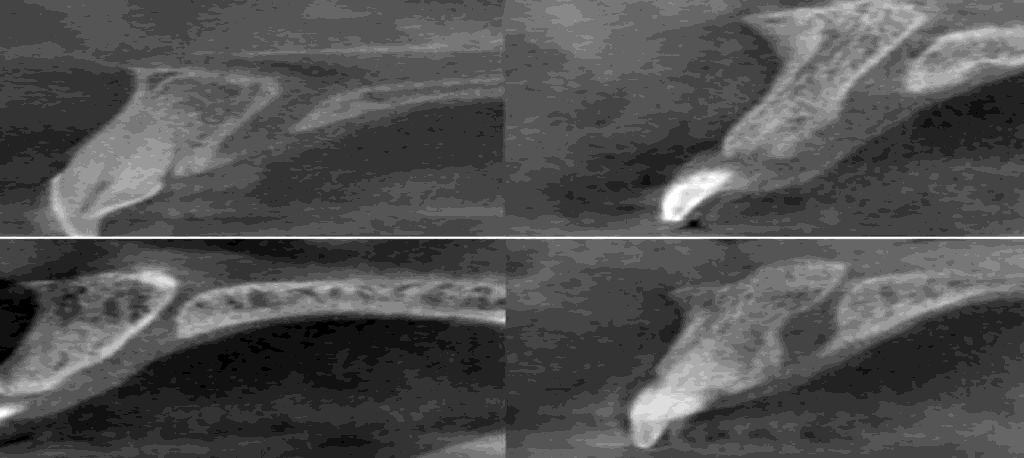
The shape and curvature of the NPC differed among cases in the sagittal view. The NPCs were thus classified into 4 categories according to their shape viewed on the sagittal sections: cylindrical, funnel, spindle and hourglass (Figure 3). The most commonly encountered shape was the cylindrical shape 22 (48.9%), and the least common was the spindle shape, seen in 3 (6.7%) subjects. Maximum cylindrical shape 11 (61.1%) was observed in age group of 36-57 years having p value 0.547. Gender wise out of 22 cylindrical shape females 8 was more common (53.3%) than male 14 (46.7%) having p value 0.235. Statistically significant differences between the genders and between the different age groups with respect to the shape of the NPC were not observed (Table 6 and Table 7).

Figure 3: Sagittal section shows the four shapes of the nasopalatine canal (cylindrical, funnel, hourglass and spindle).
| Shape of Nasopalatine canal (SNPC) | Total | ||||||
|---|---|---|---|---|---|---|---|
| 1 | 2 | 3 | 4 | ||||
| Age GRP | 14-35 yrs | Count | 8 | 5 | 3 | 3 | 19 |
| % within age GRP | 42.10% | 26.30% | 15.80% | 15.80% | 100.00% | ||
| 36-57 yrs | Count | 11 | 5 | 2 | 0 | 18 | |
| % within age GRP | 61.10% | 27.80% | 11.10% | 0.00% | 100.00% | ||
| 58-79 yrs | Count | 3 | 3 | 2 | 0 | 8 | |
| % within age GRP | 37.50% | 37.50% | 25.00% | 0.00% | 100.00% | ||
| Total | Count | 22 | 13 | 7 | 3 | 45 | |
| % within age GRP | 48.90% | 28.90% | 15.60% | 6.70% | 100.00% | ||
| Value | df | Asymp. Sig. (2-sided) | Exact Sig. (2-sided) | Exact Sig. (1-sided) | Point Probability | |
|---|---|---|---|---|---|---|
| Pearson chi-square | 5.991a | 6 | 0.424 | 0.45 | ||
| Likelihood ratio | 6.983 | 6 | 0.322 | 0.446 | ||
| Fisher's exact test | 5.06 | 0.547 | ||||
| Linear-by-linear association | .814b | 1 | 0.367 | 0.397 | 0.217 | 0.059 |
| N of valid cases | 45 |
a. 8 cells (66.7%) have expected count less than 5. The minimum expected count is .53. b. The standardized statistic is -.902.
Table 6: Nasopalatine canal.
| Shape of Nasopalatine Canal (SNPC) | Total | ||||||
|---|---|---|---|---|---|---|---|
| 1 | 2 | 3 | 4 | ||||
| Sex | Male | Count | 14 | 11 | 3 | 2 | 30 |
| % within sex | 46.70% | 36.70% | 10.00% | 6.70% | 100.00% | ||
| Female | Count | 8 | 2 | 4 | 1 | 15 | |
| % within sex | 53.30% | 13.30% | 26.70% | 6.70% | 100.00% | ||
| Total | Count | 22 | 13 | 7 | 3 | 45 | |
| % within sex | 48.90% | 28.90% | 15.60% | 6.70% | 100.00% | ||
Chi-Square Tests
| Value | df | Asymp. Sig. (2-sided) | Exact Sig. (2-sided) | Exact Sig. (1-sided) | Point Probability | |
|---|---|---|---|---|---|---|
| Pearson chi-square | 3.761a | 3 | 0.288 | 0.296 | ||
| Likelihood ratio | 3.903 | 3 | 0.272 | 0.371 | ||
| Fisher's exact test | 3.866 | 0.235 | ||||
| Linear-by-linear association | .112b | 1 | 0.738 | 0.869 | 0.428 | 0.123 |
| N of valid cases | 45 |
a. 5 cells (62.5%) have expected count less than 5. The minimum expected count is 1.00. b. The standardized statistic is .335.
Table 7: Shape of nasopalatine canal.
The NPCs were further classified according to their curvature. The nasal floor was regarded as the “horizontal” plane. 10 degree from the vertical were regarded to be “slanted,” and those whose course changed by 10 degree from vertical were regarded as “vertical” (Figure 4).

Figure 4: Sagittal section show the curvature of the nasopalatine canal (slanted, slanted-curved, vertical and vertical-curved).
Four types of NPCs based on curvature were noted: vertical, vertical-curved, slanted, and slanted curved. Most commonly, the NPC was found to be slanted 32 (71.1%), and only 4 subjects (8.9%) was found to have a slanted-curved and vertical curvature. Maximum cases 19 were observed in the age group of 14-35 yrs out of which (68.4%) was slanted curvature. From slanted type maximum cases was observed in the age group of 14-35 and 36-57 yrs having p value 1.000. Gender wise out of 32 slanted curvature female predominant 11 (73.3%) and 21 (70.0%) males having p value 0.113. Statistically significant differences between the genders and between the different age groups with respect to the shape of the NPC were not observed (Table 8 and Table 9)
| Curvature of Nasopalatine Canal (CNPC) | Total | ||||||
|---|---|---|---|---|---|---|---|
| 1 | 2 | 3 | 4 | ||||
| Age GRP | 1 | Count | 13 | 2 | 2 | 2 | 19 |
| % within age GRP | 68.40% | 10.50% | 10.50% | 10.50% | 100.00% | ||
| 2 | Count | 13 | 1 | 2 | 2 | 18 | |
| % within age GRP | 72.20% | 5.60% | 11.10% | 11.10% | 100.00% | ||
| 3 | Count | 6 | 1 | 0 | 1 | 8 | |
| % within age GRP | 75.00% | 12.50% | 0.00% | 12.50% | 100.00% | ||
| Total | Count | 32 | 4 | 4 | 5 | 45 | |
| % within age GRP | 71.10% | 8.90% | 8.90% | 11.10% | 100.00% | ||
| Value | df | Asymp. Sig. (2-sided) | Exact Sig. (2-sided) | Exact Sig. (1-sided) | Point Probability | |
|---|---|---|---|---|---|---|
| Pearson chi-square | 1.327a | 6 | 0.97 | 0.982 | ||
| Likelihood ratio | 2.046 | 6 | 0.915 | 0.982 | ||
| Fisher's exact test | 1.871 | 1 | ||||
| Linear-by-linear association | .073b | 1 | 0.787 | 0.85 | 0.439 | 0.074 |
| N of valid cases | 45 |
a. 9 cells (75.0%) have expected count less than 5. The minimum expected count is .71. b. The standardized statistic is -.270.
Table 8: NPCs were further classified according to their curvature
| Curvature of Nasopalatine Canal (CNPC) | Total | ||||||
|---|---|---|---|---|---|---|---|
| 1 | 2 | 3 | 4 | ||||
| Sex | Male | Count | 21 | 1 | 3 | 5 | 30 |
| % within sex | 70.00% | 3.30% | 10.00% | 16.70% | 100.00% | ||
| Female | Count | 11 | 3 | 1 | 0 | 15 | |
| % within sex | 73.30% | 20.00% | 6.70% | 0.00% | 100.00% | ||
| Total | Count | 32 | 4 | 4 | 5 | 45 | |
| % within sex | 71.10% | 8.90% | 8.90% | 11.10% | 100.00% | ||
Chi-Square Tests
| Value | df | Asymp. Sig. (2-sided) | Exact Sig. (2-sided) | Exact Sig. (1-sided) | Point Probability | |
|---|---|---|---|---|---|---|
| Pearson chi-square | 5.766a | 3 | 0.124 | 0.138 | ||
| Likelihood ratio | 7.105 | 3 | 0.069 | 0.127 | ||
| Fisher's exact test | 5.194 | 0.113 | ||||
| Linear-by-linear association | 1.443b | 1 | 0.23 | 0.297 | 0.148 | 0.061 |
| N of valid cases | 45 |
a. 9 cells (75.0%) have expected count less than 5. The minimum expected count is .71. b. The standardized statistic is -.270.
Table 9: NPCs were further classified according to their curvature
Angulations of the NPC
The slanting angle of the NPC was the angle measured between the floor of the nasal fossa and long axis of the NPC, which was considered to be the line joining the midpoint of the antero-posterior diameter at the nasal fossa level and the midpoint of the antero-posterior diameter at the level of the hard palate.
Overall, the slanting angle of the NPC ranged from 40 degree to 84 degree in reference to the “horizontal” plane. The mean angle for male was 62.63 (± 10.794, p value 0.212) and for female mean angle was 58.20 (± 11.632, p value 0.228). Age wise mean value for 1st age group was 63.00(± 10.530), 2nd age group mean value was 60.94 (± 12.859) and for 3rd age group 57.25 (± 8.242) p value between the group was 0.481. None of the subjects demonstrated negative values, which means that in all cases, the incisive foramen was located anterior to the nasopalatine foramina. Statistical analysis failed to show the correlation of the slanting angle of the NPC with age or gender (Tables 10-13).
Length of the NPC
As viewed on the sagittal plane, the length of the NPC was measured between the level of the nasal fossa and the level of the hard palate along the long axis of the canal. It ranged from 5.70 mm to 18.90 mm, and the mean value for male was 11.123 mm (± 3.4383, p value 0.041), for female 58.20 mm (± 11.632, p value 0.022). Age group wise mean value for 1st age group was 10.074 (± 2.8996) for 2nd age group 10.544 (± 2.7813), and for 3rd age group 11.513 (± 4.1474). Among different age groups, statistically significant differences in the length of the NPC were not observed. However, there was a statistically significant difference in the length of the canal between males and females were observed.
The mean antero-posterior diameter from the nasal fossa NPC as viewed in sagittal plane was 2.567 (± 1.5180, p value 0.780) for male and for female 2.447(± 0.9023, p value 0.742). Age wise distribution of mean for 1st age group was 2.579 (± 1.2921), 2nd age group was 2.439 (± .3303) and for 3rd age group 2.600 (± 1.6036), p value between the group was 0.939. The mean antero- posterior diameter from hard palate NPC as viewed in sagittal plane was 3.587 (± 1.2456, p value 0.319) for male and for female 3.200(± 1.1433, p value 0.308). Age wise distribution of mean for 1st age group was 3.279 (± 1.0283), 2nd age group was 3.472 (± 1.3314) and for 3rd age group 3.850 (± 1.4031), p value between the group was 0.546. The differences in the values between males and females and among the different age groups were not found to be statistically significant (Tables 10-13).
| Sex | N | Mean | Std. Deviation | Std. Error Mean | |
|---|---|---|---|---|---|
| Length of Incisive fossa (LIF) | Male | 30 | 3.863 | 1.1306 | 0.2064 |
| Female | 15 | 3.427 | 0.8964 | 0.2314 | |
| Length of Nasopalatine Canal Ant-Post (LNPCAP) | Male | 30 | 11.123 | 3.4383 | 0.6277 |
| Female | 15 | 9.307 | 1.671 | 0.4314 | |
| Length of Nasopalatine Canal Medio Lateral(nasal fossa) (LNPCML1) | Male | 30 | 2.567 | 1.518 | 0.2772 |
| Female | 15 | 2.447 | 0.9023 | 0.233 | |
| Length of Nasopalatine Canal Medio Lateral(hard palate) (LNPCML2) | Male | 30 | 3.587 | 1.2456 | 0.2274 |
| Female | 15 | 3.2 | 1.1433 | 0.2952 | |
| Angulation of Nasopalatine Canal (ANPC) | Male | 30 | 62.63 | 10.794 | 1.971 |
| Female | 15 | 58.2 | 11.632 | 3.003 |
Table 10: Angulations of the NPC.
| Levene'stest for equality of variances | t-test for equality of means | |||||||||
|---|---|---|---|---|---|---|---|---|---|---|
| F | Sig. | t | df | P value | Mean difference | Std. Error Difference | 95% Confidence interval of the difference | |||
| Lower | Upper | |||||||||
| LIF | Equal variances assumed | 0.522 | 0.474 | 1.303 | 43 | 0.2 | 0.4367 | 0.3352 | -0.2394 | 1.1127 |
| Equal variances not assumed | 1.408 | 34.57 | 0.168 | 0.4367 | 0.3101 | -0.1932 | 1.0665 | |||
| LNPCAP | Equal variances assumed | 4.122 | 0.049 | 1.928 | 43 | 0.041 | 1.8167 | 0.9424 | -0.0839 | 3.7173 |
| Equal variances not assumed | 2.385 | 42.996 | 0.022 | 1.8167 | 0.7617 | 0.2805 | 3.3528 | |||
| LNPCML1 | Equal variances assumed | 5.363 | 0.025 | 0.281 | 43 | 0.78 | 0.12 | 0.4265 | -0.7402 | 0.9802 |
| Equal variances not assumed | 0.331 | 41.521 | 0.742 | 0.12 | 0.3621 | -0.6109 | 0.8509 | |||
| LNPCML2 | Equal variances assumed | 0.03 | 0.864 | 1.008 | 43 | 0.319 | 0.3867 | 0.3837 | -0.3871 | 1.1604 |
| Equal variances not assumed | 1.038 | 30.383 | 0.308 | 0.3867 | 0.3726 | -0.374 | 1.1473 | |||
| ANPC | Equal variances assumed | 0.071 | 0.79 | 1.266 | 43 | 0.212 | 4.433 | 3.502 | -2.629 | 11.496 |
| Equal variances not assumed | 1.234 | 26.297 | 0.228 | 4.433 | 3.592 | -2.947 | 11.813 | |||
Table 11: Angulations of the NPC.
| N | Mean | Std. Deviation | Std. Error | 95% Confidence interval for mean | Minimum | Maximum | |||
|---|---|---|---|---|---|---|---|---|---|
| Lower bound | Upper bound | ||||||||
| LIF | 14-35 years | 19 | 3.547 | 0.9002 | 0.2065 | 3.113 | 3.981 | 1.5 | 5.7 |
| 36-57 years | 18 | 3.861 | 0.9166 | 0.216 | 3.405 | 4.317 | 2.7 | 6 | |
| 58-79 years | 8 | 3.8 | 1.7063 | 0.6033 | 2.374 | 5.226 | 2.2 | 7.6 | |
| Total | 45 | 3.718 | 1.0684 | 0.1593 | 3.397 | 4.039 | 1.5 | 7.6 | |
| LNPCAP | 14-35 years | 19 | 10.074 | 2.8996 | 0.6652 | 8.676 | 11.471 | 6.1 | 18.9 |
| 36-57 years | 18 | 10.544 | 2.7813 | 0.6556 | 9.161 | 11.928 | 5.7 | 17.8 | |
| 58-79 years | 8 | 11.513 | 4.1474 | 1.4663 | 8.045 | 14.98 | 6.4 | 19.1 | |
| Total | 45 | 10.518 | 3.0709 | 0.4578 | 9.595 | 11.44 | 5.7 | 19.1 | |
| LNPCML1 | 14-35 years | 19 | 2.579 | 1.2921 | 0.2964 | 1.956 | 3.202 | 0.8 | 5.3 |
| 36-57 years | 18 | 2.439 | 1.3303 | 0.3135 | 1.777 | 3.1 | 0.7 | 5.8 | |
| 58-79 yrs | 8 | 2.6 | 1.6036 | 0.5669 | 1.259 | 3.941 | 0.6 | 5.4 | |
| Total | 45 | 2.527 | 1.3346 | 0.1989 | 2.126 | 2.928 | 0.6 | 5.8 | |
| LNPCML2 | 14-35 years | 19 | 3.279 | 1.0283 | 0.2359 | 2.783 | 3.775 | 1.3 | 4.7 |
| 36-79 years | 18 | 3.472 | 1.3314 | 0.3138 | 2.81 | 4.134 | 1 | 6.1 | |
| 58-79 years | 8 | 3.85 | 1.4031 | 0.4961 | 2.677 | 5.023 | 2.2 | 5.8 | |
| Total | 45 | 3.458 | 1.2135 | 0.1809 | 3.093 | 3.822 | 1 | 6.1 | |
| ANPC | 14-35 years | 19 | 63 | 10.53 | 2.416 | 57.92 | 68.08 | 49 | 85 |
| 36-57 years | 18 | 60.94 | 12.859 | 3.031 | 54.55 | 67.34 | 40 | 84 | |
| 58-79 years | 8 | 57.25 | 8.242 | 2.914 | 50.36 | 64.14 | 42 | 68 | |
| Total | 45 | 61.16 | 11.15 | 1.662 | 57.81 | 64.51 | 40 | 85 | |
Table 12: NPC with age or gender.
| Sum of squares | Df | Mean square | F | Sig. | ||
|---|---|---|---|---|---|---|
| LIF | Between groups | 0.976 | 2 | 0.488 | 0.416 | 0.662 |
| Within groups | 49.25 | 42 | 1.173 | |||
| Total | 50.226 | 44 | ||||
| Total | 39.2 | 44 | ||||
| LNPCAP | Between groups | 11.676 | 2 | 5.838 | 0.608 | 0.549 |
| Within groups | 403.25 | 42 | 9.601 | |||
| Total | 414.926 | 44 | ||||
| Between groups | 0.234 | 2 | 0.117 | 0.063 | 0.939 | |
| LNPCML1 | Within groups | 78.134 | 42 | 1.86 | ||
| Total | 78.368 | 44 | ||||
| Between groups | 1.842 | 2 | 0.921 | 0.615 | 0.546 | |
| LNPCML2 | Within groups | 62.948 | 42 | 1.499 | ||
| Total | 64.79 | 44 | ||||
| ANPC | Between groups | 187.467 | 2 | 93.733 | 0.745 | 0.481 |
| Within groups | 5282.444 | 42 | 125.772 | |||
| Total | 5469.911 | 44 | ||||
Table 13: NPC with age or gender.
Discussion
The retrospective study indicated that the nasopalatine canal showed a great deal of variability with regard to its dimensions as well as to its morphological appearance. The present study found one to four foramina at the level of the nasal floor. This was in accordance with the previous studies by Mraiwa et al. and Thakur et al. they also reported one to four foramina at the level of the nasal floor. However, Song et al observed only two foramina in their respective studies. This variability in results could be due to sample differences and the various imaging techniques used in different studies. The mean inner diameter of the incisive foramen in the present study was ranged from 2.20 mm to 7.60 mm, and the mean value for male was 3.863 mm (± 1.1306, p value 0.200), for female 3.427 mm (± 0.8964, p value 0.168). Age group wise mean value for 1st age group was 3.547 (± 0.9002) for 2nd age group 3.861(± 0.9166), and for 3rd age group 3.800 (± 1.7063). These values were lower than those reported in the study by Mraiwa et al. [6] (4.6 mm) but comparable to those reported by Liang et al. [2] (3.4 mm). In the present study out of 45 subject 28 (62.2%) having heart shape incisive foramen was observed which was in accordance with Thakur et al.
Previous studies have reported that a cylindrically shaped NPC was most commonly observed [5,6]. In accordance with this, in our study, the cylindrical shape was found in most of the subjects. In contrast to Song et al who reported the predominance of the vertical type of NPC in their study, in present study, slanted canals were more commonly observed than vertical ones. Liang et al have previously reported the mean angulation of the NPC from the horizontal as 77.4 degree (± 8.9). In present study the slanting angle of the NPC ranged from 40 degree to 84 degree in reference to the “horizontal” plane. The mean angle for male was 62.63 (± 10.794, p value 0.212) and for female mean angle was 58.20 (± 11.632, p value 0.228). Age wise mean value for 1st age group was 63.00(± 10.530), 2nd age group mean value was 60.94 (± 12.859) and for 3rd age group 57.25 (± 8.242) p value between the group was 0.481. Song et al have reported the length of the NPC to be 12.0 mm (8.4- 15.8 mm) in maxilla, Mraiwa et al. have reported a mean length of 8.1 (±3.4) mm, and Liang et al. [2] in their study assessed the length of the NPC as 9.9 (± 2.6) mm. In the present study, the mean length of the NPC was found to be 10.08 mm (± 2.25). It ranged from 5.70 mm to 18.90 mm, and the mean value for male was 11.123 mm (± 3.4383, p value 0.041), for female 58.20 mm (± 11.632, p value 0.022). Age group wise mean value for 1st age group was 10.074 (± 2.8996) for 2nd age group 10.544 (± 2.7813), and for 3rd age group 11.513 (± 4.1474). Among different age groups, statistically significant differences in the length of the NPC were not observed. However, there was a statistically significant difference in the length of the canal between males and females were observed [7].
The mean antero-posterior diameter from the nasal fossa NPC as viewed in sagittal plane was 2.567 (± 1.5180, p value 0.780) for male and for female 2.447(± 0.9023, p value 0.742). Age wise distribution of mean for 1st age group was 2.579(± 1.2921), 2nd age group was 2.439 (± 1.3303) and for 3rd age group 2.600 (± 1.6036), p value between the group was 0.939. The mean antero- posterior diameter from hard palate NPC as viewed in sagittal plane was 3.587(± 1.2456, p value 0.319) for male and for female 3.200(± 1.1433, p value 0.308). Age wise distribution of mean for 1st age group was 3.279 (± 1.0283), 2nd age group was 3.472 (± 1.3314) and for 3rd age group 3.850 (± 1.4031), p value between the group was 0.546. In this study, statistically significant differences in the assessed parameters could not be observed among the different age groups and gender [8]. This could be attributed to the population chosen for this study. The greater length of the NPC in the males could be ascribed to the relatively larger cranio-caudal dimension of the face observed in the males as compared to the females. Similar findings have been previously reported by Liang et al.
Conclusion
In conclusion, this retrospective study highlighted the anatomic variability of several parameters such as general anatomy, size of incisive fossa, shape of incisive fossa, Number of opening of Stenson’s, size, shape, angulations, curvature of nasopalatine canal in single study and their correlation with different age group and sex of the subjects. Study also emphasizing the role of CBCT imaging and assessment of this anatomical landmark in treatment planning of this area for implant placement or assessment of pathologies in this region. The shape, curvature, and angulations of the canal and its antero-posterior dimensions are the most significant parameters for placement of implants in the maxillary incisor region. Additionally, the number of openings, medio-lateral dimensions, length of the canal, and level of its division may prove important when implants within the nasopalatine canal are being considered.
References
- Mardinger O, Namani-Sadan N, Chaushu G, Schwartz-Arad D. Morphologic changes of the nasopalatine canal related to dental implantation: a radiologic study in different degrees of absorbed maxillae. J Periodontol 2008; 79(9): 1659-62.
- Liang X, Jacobs R, Martens W, Hu Y, Adriaensens P, Quirynen M, et al. Macro- and micro-anatomical, histological and computed tomography scan characterization of the nasopalatine canal. J Clin Periodontol 2009; 36(7): 598-603.
- Kraut RA, Boyden DK. Location of incisive canal in relation to central incisor implants. Implant Dent 1998; 7(3): 221-225.
- Cavalcanti MG, Yang J, Ruprecht A, Vannier MW. Accurate linear measurements in the anterior maxilla using orthoradially reformatted spiral computed tomography. Dentomaxillofac Radiol 1999; 28(3): 137-140.
- Shear M, Speight P. Cysts of the oral and maxillofacial regions. (4th ed.). Oxford: Blackwell; 2007. p. 108-18.
- Mraiwa N, Jacobs R, Van Cleynenbreugel J, Sanderink G, Schutyser F, Suetens P, et al. The nasopalatine canal revisited using 2D and 3D CT imaging. Dentomaxillofac Radiol 2004; 33(6): 396-402.
- Thakur RA, Burde K, Guttal K, Naikmasur GV. Anatomy and morphology of the nasopalatine canal using cone-beam computed tomography. Imaging Sci Dent. 2013; 43(4): 273-281.
- Song WC, Jo DI, Lee JY, Kim JN, Hur MS, Hu KS, et al. Microanatomy of the incisive canal using three-dimensional reconstruction of microCT images: an ex vivo study. Oral Surg Oral Med Oral Pathol Oral Radiol Endod 2009; 108(4): 583-90.
Author Info
Vikash Ranjan1*, Soumendu Bikash Maiti2, Pratyush Sharma2, Vinay Kumar Mathadey2, Tasaduq Ahmad Wani2 and Garima Sharma2
1Department of Dentistry, Oral Medicine and Radiology, Maharashtra, India2Department of Oral Medicine and Radiology, Divya Jyoti College of Dental Sciences and Research, Modi, India
Received: 22-Jul-2021 Accepted: 29-Jul-2021 Published: 12-Aug-2021
