Case Report - (2020) Volume 8, Issue 1
An Isolated Chronic Gun Pellet Entrapped in Temporomandibular Joint: A Rare Case Report
Venkatesh Anehosur* and Priti Kumari
*Correspondence: Venkatesh Anehosur, Department of Oral and Maxillofacial Surgery, SDM College of Dental Sciences and Hospital, Sri Dharmasthala Manjunatheshwara University, Dharwad, Karnataka, India, Email:
Abstract
Purpose: To highlight an isolated gun pellet injury in the temporomandibular joint. Head and face are relatively common sites of gunshot injury and the isolated temporomandibular joint is uncommonly involved. This paper highlights a case of facial gunshot wound and the projectile was lodged at the neck of the condyle in the temporomandibular joint region causing pain, trismus and impairment in the mandible function. The projectile was identified and isolated to retrieve surgically via endaural approach. Postoperative strict physiotherapy schedule was followed to restore the function of the joint. Six months follow up revealed good range of functional movements without pain.
Keywords
Gunshot wound, pellet, projectile, Temporomandibular joint
Introduction
The intensity of gunshot wound is universally proportional to the severity of facial injury. It is a form of physical trauma which varies from simple isolated pellet to high velocity full blown injury. They are the second most sources of injury and death, after motor vehicle accidents. Among the various parts of body affected like abdomen, chest, upper and lower limbs, the facial and the skull base injuries are fatal and even if the patient survives, secondary deformities are challenging to treat. According to literature the majority of civilian fire arm injuries are sustained from handguns (86%), shotgun (8%), and rifles (5%). According to literature 36% deaths occur upon admission secondary to injuries to the chest, abdomen or brain. There is small percentage of deaths associated with isolated facial injuries. The head and face area is a relatively common site of gunshot injury [1]. The temporomandibular joint (TMJ) may be involved in many cases, as may important anatomic structures such as the facial nerve [2], with the potential for development of traumatic facial palsy [3]. Lesions of the temporal and marginal branches produce remarkable functional loss and must be repaired. Immediate reconstruction is indicated in cases of uncontaminated trauma. The pattern of trauma in gunshot wounds (GSWs) is extremely variable. These injuries may affect vital structures and cause difficult-to-control bleeding and proper initial management usually requires a multidisciplinary approach. GSWs often cause comminuted fractures with multiple small bone fragments [4] and may produce sequelae as serious as those caused by massive blunt impact to the same area. The purpose of this paper is to report a case of long standing 25 years old isolated gun pellet injury and the pellet being entrapped in the temporomandibular joint area causing functional impairment.
Case Report
33-year-old female patient reported to our unit with a chief complaint of pain and difficulty in opening and closing the mouth since three months. History revealed patient was apparently alright three months back after which she developed pain over left ear region for which she received treated unsuccessfully for ear infection. History of domestic violence 25 years back was recorded. Patient also gave history of accidental gunshot injury when she was 8 years old. Pain which is sudden onset, intermittent type and mild in nature with high in intensity with associated headache. Patient noticed pain getting aggravated on mastication and temporary relief while taking analgesic. No other associated symptoms noted. On clinical examination mild deviation of jaw towards left side while opening mouth (Figure 1). Clicking sound and crepitus noted on opening and closing of mouth over left TMJ region. Lateral excursion movement was found painful and limited towards right side. Tenderness noted over left masseter and pterygoid region. Trismus was evident with mouth opening of 2 centimeters. Provisional diagnosis of internal derangement was made and conventional radiographs orthopantamogarm and TMJ views with open and closed mouth were asked which revealed presence of a radiopaque foreign body in the left TMJ region (Figure 2). Computed tomography showed exact location of foreign body at the level of lateral to neck of condyle (Figure 3). Patient was explained about the same and re-examined for presence of entry wound which was not evident. Planned exploratory surgery under general anaesthesia was explained to the patient and consented for possible complications especially facial nerve weakness.
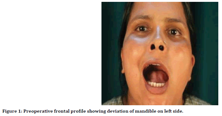
Figure 1. Preoperative frontal profile showing deviation of mandible on left side.
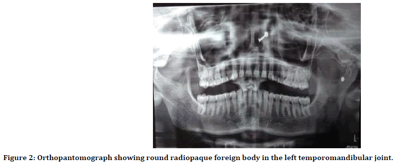
Figure 2. Orthopantomograph showing round radiopaque foreign body in the left temporomandibular joint.
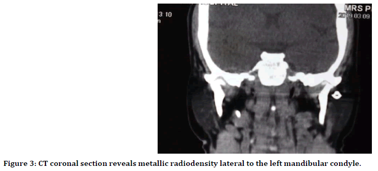
Figure 3. CT coronal section reveals metallic radiodensity lateral to the left mandibular condyle.
After adequate patient preparation with routine blood investigations the foreign body was retrieved by endaural surgical approach under general anaesthesia (Figure 4). The incision is made behind the prominence of the tragus. The skin flap is reflected over the cartilage of the tragus, and then, blunt dissection is continued along the cartilage of external auditory canal to reach the root of zygomatic arch. The dissection carried through the superficial temporal fascia to prevent damage to facial nerve branch. Subperiosteal dissection was carried out on the arch to expose the condyle and neck. There was evidence of tissue adhesion and discoloration of soft tissue helped in locating a round metallic like structure measuring about 0.5 X 0.5 mm (Figure 5). The projectile was removed in to and the surgical site was cleaned with saline solution (Figure 6). Intraoperatively 4 cms of mouthoepening was achieved with exercise regimen. The surgical site was closed in layers with minivac drain. The postoperative check radiograph was done (Figure 7) healing process uneventful, with good resolution of the initial symptoms. With a follow- up of 1 month, the patient is in good health with a normal range of temporomandibular joint movement.
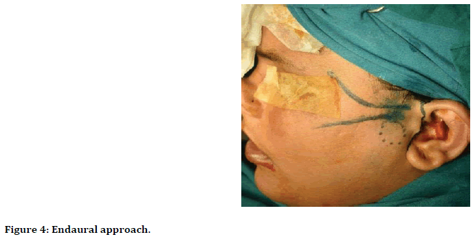
Figure 4. Endaural approach.
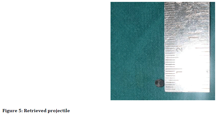
Figure 5. Retrieved projectile
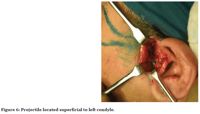
Figure 6. Projectile located superficial to left condyle.
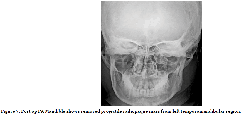
Figure 7. Post op PA Mandible shows removed projectile radiopaque mass from left temporomandibular region.
Discussion
Oral and maxillofacial gunshot injuries are usually fatal due to close proximity with vital structures. Here, we report a case in which radiographic evidence of foreign bodies in the left temporomandibular region exposed a history of a gunshot injury. The patient did not have any major complaints except for pain and reduced mouth opening. Gunshot wounds, when they are not fatal, can cause severe organ damage [5]. The degree of damage caused by a bullet depends on the type of bullet, the shooting distance, the initial velocity, the direct or indirect nature of the impact and the shooter’s intention [6]. Incarceration of a bullet with in TMJ is due to either a low velocity bullet, or a high velocity bullet shot from a long distance or slowed by intermediate impacts, as in our patient. Bullet impacting on the face predominantly lodge in the maxillary, frontal, ethmoid and TMJ, in decreasing order of frequency. Some bullet made up to toxic metal which can damage the soft tissue as well as hard tissue like lead containing shotgun pellet after penetrated in head and neck region [7]. Thus, the late manifestation of foreign body materials should not be underestimated. According to a case report by Claros, et al. a patient developed chorioretinitis sclopetaria due to foreign body embedded in infraorbital region for 22 years [8].
Infraorbital foreign bodies can cause proliferative chorioretinitis responsible for irreversible lesion. The diagnosis of TMJ foreign body was accidently detected on the basis of the history of the accident and clinical examination and it was confirmed by conventional radiographs showing a radiopaque foreign body. Computed tomography defines the exact position and anatomical relations of the foreign body. The role of Magnetic Resonance Imaging (MRI) is highlighted in especially in neck region, when the foreign body is close proximity to the important vessels of the neck [9]. When a foreign body is inaccessible or lodged close to a vital organ, conservative management is generally recommended as surgical intervention could be more fatal than lodged pellet [10]. However, surgery may be decided after evaluation of several parameters’ accessibility, the organic or inorganic nature of the foreign body anatomical relation with neurovascular bundle, branch of facial nerve auriculotemporal nerve, superficial temporal artery [11]. In the present case, the patient was advised for exploratory surgery due to the presence of symptoms and the accessibility of the pellet and it would benefit the patient psychologically.
Though there are many approaches for TMJ, the endaural approach for removal of a foreign body was opted over preauricular as surgical access required was minimal and because of its cosmesis. In conclusion, we report a very rare case of long standing gun pellet entrapped in TMJ region causing symptoms and entailing surgical exploration to retrieve the same with nil functional morbidity.
Acknowledgment
Authors would like to thank Dr Niranjan Kumar, Vice Chancellor, SDM University, Medical Director and Director of SDM Craniofacial Centre and Dr Srinath Thakur, principal SDM College of dental sciences and hospital for the support, encouragement and facilities provided.
Funding
Nil funding received.
Competing Interest
Nil conflict of interest.
Ethical Approval
The manuscript was cleared by Institutional review board for publication.
Patient Consent
Consent has been taken.
References
- Kara MI, Polat HB, Ay S. Penetrated shotgun pellets: A rare case report. Eur J Dent 2008; 2:51-62.
- Hollier L, Grantcharova EP, Kattash M. Facial gunshot wounds: A 4-year experience. J Oral Maxillofac Surg 2001; 59:277-282.
- Kiran DN, Mittal S. Gunshot (Pellet) injury to the maxillofacial complex: A case report. Chin J Traumatol 2014; 17:170-172.
- Malik FR, Singh SK, Madeshiya S, et al. Evaluation of complete profile and outcome of gunshot injuries in tertiary care centre. Int Surg J 2019; 6:397-402.
- Pires MSM, Giongo CC, Antonello GM, et al. An interesting case of gunshot injury to the temporomandibular joint. Craniomaxillofac Trauma Reconstr 2015; 8:79-82.
- Ongom PA, Kijjambu SC, Jombwe J. Atypical gunshot injury to the right side of the face with the bullet lodged in the carotid sheath: A case report. J Med Case Reports 2014 8:29.
- Cunningham LL, Haug RH, Ford J. Firearm injuries to the maxillofacial region: An Overview of current thoughts regarding demographics, pathophysiology, and management. J Oral Maxillofac Surg 2003; 61:932-942.
- Claros P, Claros A. Intraorbital foreign body: A rifle bullet removed 20 years after the accident. J European Annals Otorhino Head Neck Disease 2017; 134:63-65.
- Patini R, Arrica M, Di Stasio E, et al. The use of magnetic resonance imaging in the evaluation of upper airway structures in paediatric obstructive sleep apnoea syndrome: A systematic review and meta-analysis. Dentomaxillofac Radiol 2016; 45:20160136.
- Munishwar PD, Gupta A, Bajantri A, et al. Ballistic trauma. J Ind Acad Oral Med Radiol 2016; 28:184-187.
- Godhi S, Mittal GS, Kukreja P. Gunshot injury in the neck with an atypical bullet trajectory. J Maxillofac Oral Surg 2011; 10:80-84.
Author Info
Venkatesh Anehosur* and Priti Kumari
Department of Oral and Maxillofacial Surgery, SDM College of Dental Sciences and Hospital, Sri Dharmasthala Manjunatheshwara University, Dharwad, Karnataka, IndiaCitation: Venkatesh Anehosur, Priti Kumari, An Isolated Chronic Gun Pellet Entrapped in Temporomandibular Joint: A Rare Case Report, J Res Med Dent Sci, 2020, 8(1): 162-166.
Received: 25-Oct-2020 Accepted: 05-Feb-2020
