Research - (2024) Volume 12, Issue 8
G. Karthika*, M.K Rajasekar, Vandana Arun and Rajeshwari Nachiyar
*Correspondence: G. Karthika, Department of Otorhinolaryngology, Sree Balaji medical college and hospital, India, Email:
Abstract
Adenoameloblastoma, also known as adenomatoid odontogenic tumor (AOT), is a rare benign tumor sometimes confused for other cystic lesions owing to its clinical and radiological similarities. A 23-year-old man appeared with a painless facial swelling that was first identified as a nasolabial cyst. However, histological examination after surgical removal showed the tumor to be an adenoameloblastoma. Using the Caldwell-Luc technique, the tumor was entirely removed. Over three years, the patient recovered uneventfully and had no recurrence. This case highlights the need to include adenoameloblastoma in the differential diagnosis of maxillofacial swellings and the need for histological confirmation for correct diagnosis and therapy.
Keywords
Adenoameloblastoma, Adenomatoid Odontogenic Tumor (AOT), Nasolabial cyst, Maxillary swelling, surgical excision.
Introduction
Adenoameloblastoma, often referred to as Adenomatoid Odontogenic Tumor (AOT), is a rare benign tumor that originates from the outer layer of cells in the teeth. It is most usually seen in young people, with the majority of instances occurring within the second decade of life. With a preference for the anterior maxilla, it accounts for 3-7% of all odontogenic tumors. It often manifests in conjunction with an unerupted tooth, especially a canine [1, 2]. Ameloblastic Odontoma (AOT) is often without symptoms and it is frequently detected by chance during regular dental scans or as a result of a gradually growing, painless swelling in the maxillofacial area. On radiographs, the appearance of AOT is usually a well-defined, unilocular radiolucent abnormality that may include areas of calcification. It may frequently resemble other cysts or tumors that originate from the teeth, such as dentigerous cysts or calcifying odontogenic cysts [3]. The resemblance seen in the radiographic images might create difficulties in distinguishing between various diagnoses, potentially resulting in incorrect identification, as shown in this particular example where the abnormality was first mistaken for a nasolabial cyst. AOT is histologically characterized by the presence of unique duct-like structures, which are often encircled by spindle-shaped or cuboidal epithelial cells grouped in rosettes or nests. Calcifications inside the tumor are a distinctive characteristic that aids in differentiating it from other odontogenic tumors [4]. Accurate diagnosis is crucial in providing appropriate therapy and averting unnecessary treatment, despite the benign nature and low recurrence risk of AOT after surgery [5]. This case highlights the significance of considering AOT (Adenomatoid Odontogenic Tumor) as a potential cause of maxillary swellings, especially in younger individuals who exhibit unusual clinical characteristics.
Case Report
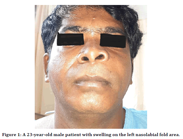
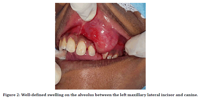
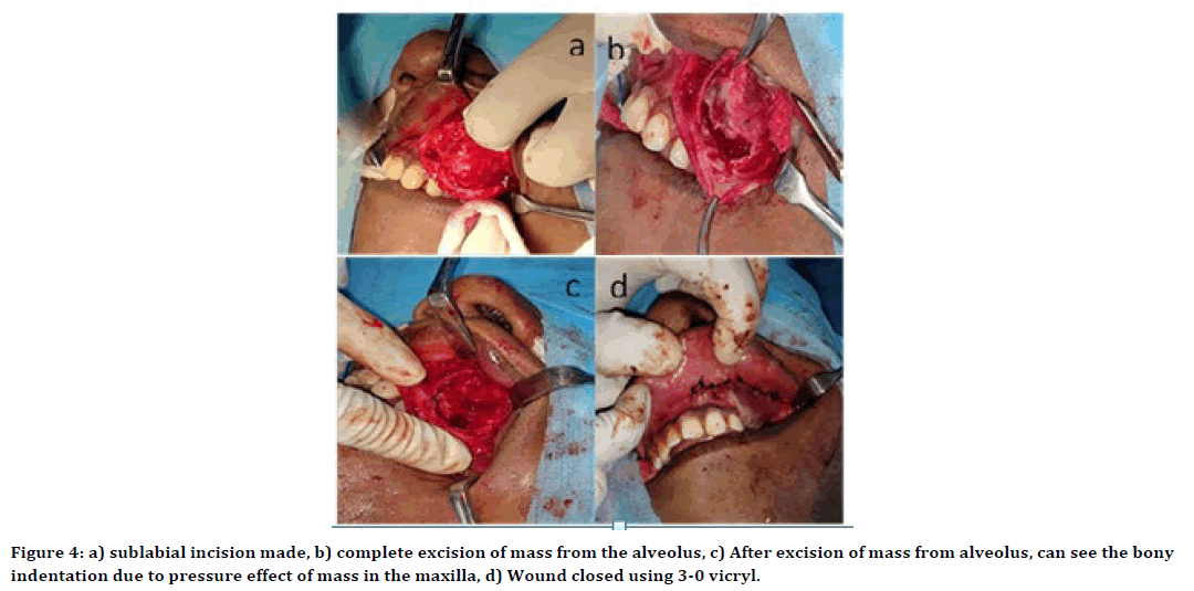
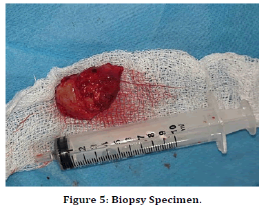
This case report describes a rare instance of adenoameloblastoma in the maxilla, which was first misdiagnosed as a nasolabial cyst. A 23-year-old male patient presented to the Outpatient Department (OPD) with a main complaint of an asymptomatic lump located on the left side of his face, especially below the nose and in the upper left tooth area. For over a year, this mass has been there. The patient had no previous record of face injury, and the swelling originally presented as a little bulge, about the size of a pea. Over time, the bulk progressively became larger. The patient did not report any concomitant symptoms such as pain or discomfort. Facial asymmetry was seen during extra oral examination owing to swelling in the left nasolabial fold area (Figure 1). A single well-defined enlargement of about 3 cm x 2 cm was seen intraorally on the alveolus between the left maxillary lateral incisor and canine (Figure 2). The mass-produced minor labiopalatal enlargement of the maxillary alveolus and damaged the labial vestibule. Notably, the adjacent teeth had been displaced by the swelling. The swelling had a smooth surface, with mucosa covering it normally, and had a solid to hard firmness. There was no visible or palpable pulse, and the mass was not painful when touched. A fine needle aspiration was conducted, but no fluid was obtained. Computed Tomography (CT) scans showed indications of cortical bone loss in addition to well-demarcated hypodense lesions with scattered hyper dense foci (Figure 3). These observations led to a differential diagnosis, taking into account the potential presence of either a nasolabial cyst or an odontogenic cyst. The patient had surgical intervention using the Caldwell Luc technique. The growth was completely eliminated by enucleation, and the sample was submitted for histological analysis. The intraoperative scans revealed a bony indentation in the maxilla and the full removal of the tumor from the location. After excision, gel foam was used to fill the cavity left behind, and 3-0 Vicryl sutures were used to seal the incision (Figure 4). Upon gross examination, the specimen exhibited a grey-white globular form measuring 3.5 cm x 2 cm x 1 cm, with an exterior surface displaying a tint ranging from grey-white to tan (Figure 5). The cyst wall had a thickness of 0.2 cm, and the cut surface displayed patches that ranged from grey-white to grey-brown. There were no visible solid regions or papillary excrescences. Under a microscope (Figure 6), a well-defined tumor consisting of tumor cell strands with one to two cell thickness layers around the periphery was seen. There were sheets of tumor cells with stellate shapes and a cribriform arrangement in the central part of the lesion. Globular masses of calcification were found. There were also columnar tumor cells with luminal basophilic secretions at the lesion's central area. Crucially, no necrosis or mitosis was seen. The sections taken from the removed margin showed no significant histological abnormalities. The histological results confirmed the presence of an adenomatoid odontogenic tumor, which is also referred to as adenoameloblastoma. Throughout the three-year follow-up, the patient's surgical recovery was uncomplicated and there was no sign of recurrence.

Figure 1. A 23-year-old male patient with swelling on the left nasolabial fold area.

Figure 2. Well-defined swelling on the alveolus between the left maxillary lateral incisor and canine.

Figure 3. CT image showing well-demarcated hypodense lesion with scattered hyperdense foci in both axial and coronal cut.

Figure 4. a) sublabial incision made, b) complete excision of mass from the alveolus, c) After excision of mass from alveolus, can see the bony indentation due to pressure effect of mass in the maxilla, d) Wound closed using 3-0 vicryl.

Figure 5. Biopsy Specimen.

Figure 6. Microscopy demonstrating whorls of epithelial islands with calcification areas, ductal and sheets of tumor cells with stellate shapes and a cribriform arrangement in the central part of the lesion surrounded by a well-defined mature capsule.
Discussion
Adenoameloblastoma, also referred to as an adenomatoid odontogenic tumor (AOT), is a rarely occurring benign tumor that predominantly affects the maxilla and is frequently associated with unerupted teeth, especially among young adults. The clinical appearance of this condition often involves painless swelling and facial asymmetry. However, due to its rarity and similarity to other cystic lesions like nasolabial cysts, it may easily be misdiagnosed. This instance highlights the difficulty in diagnosing adenoameloblastoma since it may resemble more typical maxillofacial lesions. The patient's manifestation of a painless, gradually expanding mass on the left side of the face, which was first identified as a nasolabial cyst, exemplifies the intricate nature of diagnosing adenoameloblastoma. Nasolabial cysts, which are often seen, usually manifest as soft, fluctuating swellings in the vicinity of the nasolabial fold. On the other hand, the lesion in this patient's tooth displacement and hard consistency are more suggestive of an odontogenic origin [6]. These specific symptoms should lead to considering other possible diagnoses, such as odontogenic tumours like adenoameloblastoma. An essential part of the first assessment is radiographic examination; radiopaque foci and well-circumscribed radiolucencies are common features of adenoameloblastomas. These characteristics were obvious in this case, where the CT scan indicated hypodense lesions with scattered hyperdense patches and loss of cortical bone. However, the similarity in radiographic characteristics between adenoameloblastomas and other cystic tumours makes the diagnosis procedure more difficult, requiring confirmation by histological examination [7]. Histopathologically, adenoameloblastomas have unique features that set them apart from other odontogenic tumors. This tumor is characterized by the presence of stellate-shaped tumor cells grouped in a cribriform pattern with basaloid cells forming anastomosing threads. Furthermore, the identification of calcifications and the lack of mitotic activity or necrosis provide further evidence for the diagnosis. Differentiating adenoameloblastoma from other cystic lesions and benign tumors, which may have less distinct or more aggressive characteristics, requires an understanding of these histological findings [8]. Due to the benign nature of adenoameloblastoma and its low recurrence incidence, surgical excision is still the preferred course of therapy. To limit the chance of tumor regrowth, it is crucial to completely remove the tumor, since inadequate excision has been linked to its recurrence in some instances [9]. The use of the Caldwell Luc technique in this particular case facilitated efficient entry to the maxillary lesion and accomplished the complete removal of the tumor. The patient's smooth and uncomplicated recovery after surgery, together with the absence of any recurrence throughout three years of observation, highlights the efficacy of this method. The present case emphasizes the need to take into account uncommon odontogenic tumors when differentiating the diagnosis of maxillofacial swellings, especially when the clinical and radiographic characteristics are unclear. Prompt and precise diagnosis is essential for assuring proper surgical treatment and avoiding the return of the condition. Furthermore, the resemblance in how adenoameloblastoma and more prevalent cystic lesions are presented highlights the need for a comprehensive approach that includes clinical, radiographic, and histological assessments [10].
Conclusion
The differential diagnosis of maxillary swellings should include adenoameloblastoma, an uncommon but serious tumor, particularly in young individuals with unusual symptoms. To get good results and minimize the danger of recurrence, it is crucial to have an accurate diagnosis and to perform full surgical excision.
References
- Nevil BW, Damm DD, Allen CM, et al. Oral and Maxillofacial Pathology 4th ed. philadelphia: WB.
- Puneet Batra BD. Adenomatoid odontogenic tumour: review and case report. J Can Dent Assoc. 2005; 71:250-3.
- John JB, John RR, Maroli RK. Adenomatoid odontogenic tumor: A report of a case and review of literature. Indian J Dent Res. 2017; 28:70-74.
- Deepa DS, Bal R, Choudhary K. Adenomatoid odontogenic tumor: A clinicopathologic correlation. J Oral Maxillofac Pathol. 2019; 23:445-448.
- Sarode SC, Sarode GS, Waknis P, et al. Adenomatoid odontogenic tumor: A review of unusual clinical and histological features. World J Clin Cases. 2019; 7:1022-1031.
- Rajendran R. Shafer's textbook of oral pathology. Elsevier India; 2009.
- Zhang J, Wang H, He X, et al. Adenomatoid odontogenic tumor: A clinicopathological, radiographic, and immunohistochemical study of 37 cases. Head Neck Pathol. 2020; 14:123-129.
- El-Naggar AK, Chan JK, Rubin Grandis J, et al. WHO classification of head and neck tumours. (No Title). 2017.
- Liu J, Li C, Zhang L, et al. Association of tumour-associated macrophages with cancer cell EMT, invasion, and metastasis of Kazakh oesophageal squamous cell cancer. Diagnostic pathology. 2019; 14:1-9.
- Kamboj M, Juneja M, Wadhwan V. Adenomatoid odontogenic tumor: A rare case report and literature review. J Oral Maxillofac Pathol. 2018; 22(Suppl 1):S87-S91.
Indexed at, Google Scholar, Cross Ref
Indexed at, Google Scholar, Cross Ref
Author Info
G. Karthika*, M.K Rajasekar, Vandana Arun and Rajeshwari Nachiyar
Department of Otorhinolaryngology, Sree Balaji medical college and hospital, IndiaReceived: 29-Jul-2024 Published: 27-Aug-2024
