Research - (2020) Volume 8, Issue 3
A Survey of Knowledge, Practices and Mishaps in Relation to Post Placement for Endodontically Treated Teeth
Mohammed Zahran1, Mohammed Tharwat Hamed2*, Ghada Naguib3, Dania Sabbahi4, Rawan Tayeb Department of Dental Public Health5 and Hisham Mously6
*Correspondence: Mohammed Tharwat Hamed, Department of Fixed Prosthodontics, Cairo University School of Dentistry, Cairo, Egypt, Email:
Abstract
Background: In the clinical settings, post and core system is commonly used as a restorative tooth treatment. However, various cases of failure have been documented concerning its failure of endodontically treated teeth.
Objective: The study aims to examine the level of knowledge concerning the post and core failure reasons among individuals in Saudi Arabia. It intends to assess the failures or mistakes which occur during post space preparation, post-placement, or after final coronal restoration in KAU.
Methods: A cross-sectional study design has been used following a quantitative approach. The questionnaire was distributed among 200 individuals (dental students, interns, general practitioners, and residents) randomly, which was then analyzed statistically.
Results: Findings reported the use of rubber dam, sodium hypochlorite as irrigation type, management through MTA (Mineral trioxide aggregate) repair and follow-up. It also showed that the failure of the restorative teeth depends on the tooth size, type, time elapsed, and repair material.
Conclusion: The study concludes that the knowledge level concerning the post and core restorative system differs for the study population. It emphasizes that standardized guideline must be set for enhancing the knowledge and improving the restorative techniques. This also helps in preventing the related mishaps.
Keywords
Failure of post and core system, Post and core system, Restorative treatment, Saudi Arabia
Introduction
In the contemporary restorative dentistry, the endodontic teeth reconstruction is challenging given the loss of teeth either totally or partially by erosion, caries, abrasion, trauma, previous restorations, or endodontic access. Yee et al. outlined that in the United States (US) alone, about 15 million individuals are receiving root canal treatment [1]. Concerning the restoration strategies, Alenzi et al. have emphasized that various parameters affect the prognosis of endodontically treated teeth (ETT) such as its final restoration type, the design of the post and core material, remaining tooth structure and ferrule presence [2]. Bakirtzoglou et al. highlighted that the complex of post and core maps out the foundation for the definitive restoration, which must be consistent with the fundamental principles to help in delivering coverage restoration [3,4]. Most studies confirm that the post and core system have increased the survival potency of restored maxillary anterior teeth from 82 to 96 percent (comprising a six to ten years follow-up period) [5]. Few studies have demonstrated that providing post-treatment may not be necessary for all root-filled teeth [6,7]. It has been found that the core restoration also provides the same result as that for the post restorations, particularly when the coronal tissue loss in teeth is minimum [8,9].
Despite this, the post and core system has been documented to increase the tooth restoration complication [10]. Santos-Filho et al. clarify that an abutment root may only be weakened by post when it exerts force on the roots [11]. The most adverse effect is observed on the anterior teeth when it is subjected to non-axial loads of 135o [3]. Generally, the subjected system should demonstrate resistance only when the occlusal load is higher than the average during function [12]. The post length also determines the retention as well as the resistance of the tooth, such as an appropriate length of post mitigate the applied stress on the tooth, which helps in better stress dissemination [13]. An earlier ten-year retrospective study has reported post dislodgement as the most frequent complication that might face the post and core system [14].
One of the prime concerns raised by dentists is the occurrence of coronal microleakage. In case, a tooth has been inadequately restored, the microorganism of root canal filing may get into the root canal, which stresses a well-sealed restoration to mitigate the reactivation of the dormant microorganisms [15]. Multiple studies reported that there is no significant difference in leakage of the apical seal after immediate or delayed post space preparation while other studies stated the opposite [16,17].
Post space preparation requires the removal of gutta-percha, which can be achieved by different techniques (mechanical, thermal, or chemical) [18]. Kuzekanani compared the effects of the heat system and two types of rotary drills to analyze the minimal dye leakage between these three groups. It found that gutta-percha removal is clinically insignificant, giving the residual amount of gutta-percha. Chemical removal of gutta-percha can be achieved using solvent, such as oil of eucalyptus and chloroform. However, some of these materials, and especially chloroform, are hazardous to use, as they are toxic and potentially carcinogenic. Other solvents lead to a dimensional change in the gutta-percha, leading to increased micro-leakage.
Concerning the post material type, several studies reported that root fractures occur more frequently with metal posts than with fiber posts. Also, higher tooth structure loss observed when metal post systems were placed. Several surveys evaluated the knowledge and practices of practitioners for restoring ETT in several countries: Switzerland, [19] Saudi Arabia, [15,20,21] India [22] and Germany [23]. Most of these surveys focused on the knowledge and practices of practitioners about restorations of ETT without reporting the mishaps that might occur during the restoration of ETT using different post systems. Therefore, this study was conducted to assess the prevalence of mishaps and iatrogenic errors during post space preparation and post placement among undergraduate students, interns, newly graduated clinicians and residents at Faculty of Dentistry, King Abdulaziz University (KAU) in Jeddah, Saudi Arabia with a special focus on the nature of these mishaps and their common causes.
Materials and Methods
Research design
A descriptive cross-sectional study design was adopted for assessing the practices followed by participants during post space preparation and post-placement using a quantitative approach. The selection of the study design is based on its efficacy to produce holistic results and provide new insights on the topic in an unbiased way [24]. On the other hand, this was also found parallel with the other studies that have been conducted in a similar discipline [21].
Research setting
The study was conducted in the Faculty of Dentistry and University Dental Hospital at KAU. The research population was inclusive of dental students, interns, general practitioners, and residents as it helps in overcoming the gap that prevailed in the earlier studies [15,20,21].
Recruited participants
The participants recruited comprised of 4th and 5th year dental students, interns, general practitioners, and residents. Initially, 289 participants were invited, though only 200 individuals showed their willingness, with a response rate of (69.5%).
Ethical consideration
Prior to the study, the researcher obtained approval from the Research Ethics Committee at Faculty of Dentistry, KAU. The study scope and its objectives were communicated to the participants, following which the participant consent was attained electronically. The researcher also communicated the anonymity and confidentiality of the data, while also highlighting participants right to withdraw from the study at any time.
Data collection
The data was collected through a survey using a close-ended questionnaire. The questionnaire was distributed online, and a period of 6 weeks was provided for completing the questionnaire. The questionnaire comprised of three parts, where first part collected the demographic data, second part collected data pertaining to the number of placed posts, isolation measures, time of post space preparation, factors of canal selection for post-placement, removal of gutta-percha and post cementation. The third part collected data concerning the post space preparation mishaps, patientrelated information, time of incidence, type of incidence, tooth-related information, and its management. Prior to questionnaire distribution, the face validity was evaluated by three experts, who reviewed the questionnaire and suggested improvements.
Data analysis
The collected data were statistically analyzed using IBM Statistical Package for Social Science for Windows (Version 23, SPSS Inc., IBM, Somers, New York, USA) Descriptive analysis was conducted, which provided results in the form of frequencies and percentages.
Results
Two hundred questionnaires were filled. More than half of the participants were females (54%). Whereas, it also showed that most of the sample were interns and sixth-year students (87%). While remaining sample (13%) belonged to the population of general practitioners and residents at Faculty of Dentistry (KAU).
Practices related to post placement
Number of placed posts within last year
Majority of participants (72.5%) reported that they had done less than five fiber posts and cores, and 57.5% had done less than five casted posts and cores (Table 1).
| Number of post last year | Fiber post (%) | Cast post (%) |
|---|---|---|
| None | 9.5 | 37.5 |
| Less than 5 | 72.5 | 57.5 |
| Between 5 to 10 | 14.5 | 2 |
| Between 11 to 20 | 2 | 2 |
| More than 20 | 1.5 | 1 |
Table 1: Number of placed posts within last year.
Practices related to isolation, root canal irrigation and post disinfection
Concerning the rubber dam, the participants showed that only 32% are always using rubber dam isolation, while 16% had never used a rubber dam during post placement on procedural bases (Table 2). Similarly, participants were asked about the type of irrigation solution after guttapercha removal during post space preparation, which revealed that 47% of them were using sodium hypochlorite, while hydrogen peroxide was used by 0.5% of participants (Table 2). When asked if they tend to disinfect the post (using appropriate disinfectant, e.g. Sodium hypochlorite or Iodophors) before trying it, 38% highlighted that they always tend to disinfect, while 6% answered that they rarely used disinfectant for posts. When asked how often they place the post immediately root canal obturation, 26% of participants stated that they mostly tend to do that, 22% others said that they had never done such thing.
| Percentage | |
| How often do you use rubber dam during post placement? | |
| Never | 16 |
| Rarely | 7 |
| Sometimes | 20 |
| Mostly | 34 |
| Always | 33 |
| Irrigation type (after gutta-percha removal) | |
| Sodium Hypochlorite | 47 |
| Saline | 26 |
| EDTA | 3.5 |
| Hydrogen Peroxide | 0.5 |
| None | 23 |
| How often do you disinfect the post before trying it? | |
| Never | 27 |
| Rarely | 5 |
| Sometimes | 15 |
| Mostly | 16 |
| Always | 38 |
| How often do you place the post (if needed) immediately after endodontic treatment? | |
| Never | 22 |
| Rarely | 19 |
| Sometimes | 25 |
| Mostly | 26 |
| Always | 9 |
Table 2: Practices followed by participants during post placement (n=200).
Factors guiding the canal selection for postplacement in multi-rooted teeth
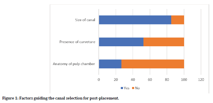
Seventy-five percent of the participants indicated that size of the canal are among the factors that they considered for selecting the canal for post placement, while about 53% and 26% reported that presence of curvature in the canal and pulpal anatomy are among the factors to be considered, respectively (Figure 1).

Figure 1. Factors guiding the canal selection for post-placement.
Removal of gutta-percha
Following of post space preparation; guttapercha removal could be established by different techniques, 76% of participants stated that they are using rotary systems for guttapercha removal. Order of burs for gutta-percha removal can be done from smallest to largest, or from largest to smallest. However, 76% of participants started from smallest to largest. While removing gutta-percha from the root canal, part of gutta-percha could be left in the canal, 99% of participants reported that they tend to leave 3-5mm apically. After gutta-percha removal, 85% of participants reported that they are taking radiographs to ensure the quality of work and self-evaluation (Table 3).
| Percentage | ||
| Method for Gutta-Percha removal from the canal | ||
| Heat | 20% | |
| Rotary | 76% | |
| Chemical | 5% | |
| Order to be followed when using Gates Glidden for gutta-percha removal | ||
| Smallest first | 76% | |
| Largest first | 16% | |
| Doesn’t matter | 4% | |
| I don’t know | 6% | |
| Remaining of gutta-percha to be left in the canal | ||
| 1-2 mm | 2% | |
| 3-5mm | 99% | |
| All GP | 0% | |
| How often do you use radiograph for verification after gutta-percha removal? | ||
| Never | 1% | |
| Rarely | 1% | |
| Sometimes | 4% | |
| Mostly | 11% | |
| Always | 85% | |
Table 3: Practices followed by participants during gutta-percha removal.
Cement delivery
Delivering of cement before post placement could be done by different techniques and instruments, 58% of participants were using special tips for cement delivery (Table 4). During cement delivery, 52% of participants deliver the cement up to full length of the canal.
| Percentage | |
| How do you deliver the cement in the canal? | |
| Special Tip | 58% |
| Lentulo Tip | 14% |
| K File | 18% |
| Paper Point | 10% |
| Which proportion of canal do you fill with the cement? | |
| Fourth of Canal | 5% |
| Half of Canal | 9% |
| Sixth of Canal | 16% |
| Full Length | 53% |
| I don’t know | 17% |
Table 4: Practices followed by participants during post cementation.
Post space preparation mishaps
Incidence of mishaps reporting
Fifty-seven participants (28.5%) of the sample reported incidences related to post placement. These incidences occurred in 18 to 35 years patients in 77% of the cases. About 33% of these incidences were during gutta-percha removal, while 30% were during drilling using different posts systems, 30% were during post cementation, and 7% during impression making for the post. Strip perforation was the most common complication reported for 33% of the cases and rotatory systems were used to remove the gutta-percha in 63.2% of the cases.
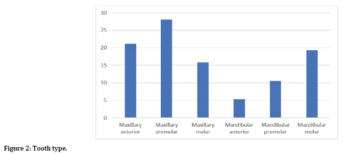
Maxillary teeth (64%) were mostly affected teeth by mishaps as shown in Figure 2. Seventyseven percent of the involved teeth were endodontically re-treated and the rubber dam was used at the time of incidence in 63% of cases. Students performed root canal treatment in about 74% of the cases. Majority of cases were not compromised as 54% of the cases had 3 or 4 remaining walls after caries excavation.

Figure 2. Tooth type.
About 63% of the practitioners tend to manage their cases by themselves and about 63% of them follow-up their cases after the incidence. Mineral trioxide aggregate (MTA) was the solution for the perforations in about 44% of the cases. Only 5 mishaps were managed by extraction. Three of these cases were perforations and 2 cases were due to periodontal complications after excessive heat application. Please refer to (Table 5) for more details about these incidences.
| Percentage | |
| Age of the patient | |
| 18-25 years | 36.8 |
| 26-35 years | 40.4 |
| 36-45 years | 5.3 |
| More than 46 years | 17.5 |
| When did the incidence occur? | |
| During gutta percha removal | 33.3 |
| During post space preparation (rotary) | 29.8 |
| During post space impression making | 7 |
| During post try-in or cementation | 29.8 |
| What was the incidence? | |
| Strip perforation | 31.6 |
| Perforation | 14 |
| Post fracture | 5.3 |
| Post dislodgment | 10.5 |
| Periodontal complication | 3.5 |
| Remove of all the gutta percha | 8.8 |
| Issues related to post seating or fit | 12.3 |
| Ledge | 3.5 |
| Others | 10.7 |
| What instrument/ technique was used for gutta percha removal? | |
| Heat | 29.8 |
| Rotary | 63.2 |
| Chemicals | 7 |
| Was the tooth endodontically re-treated? | |
| Yes | 77.2 |
| No | 22.8 |
| Were rubber dam used at time of incidence? | |
| Yes | 63.2 |
| No | 36.8 |
| Who performed the root canal treatment? | |
| Student | 73.7 |
| General Dentist | 21.1 |
| Specialist | 5.3 |
| How many walls remained after caries excavation? | |
| 1 wall | 10.5 |
| 2 walls | 35.1 |
| 3 walls | 43.9 |
| 4 walls | 10.5 |
| Did you manage the case? | |
| Yes | 63.2 |
| No | 10.5 |
| Referred to specialist | 26.3 |
| How did you manage? | |
| Repairing it with MTA | 43.9 |
| Extracting the involved tooth | 8.8 |
| Re-obturating the canal | 8.8 |
| Recement the post | 7 |
| Leave it as it is | 14 |
| Other | 17.5 |
| Did you read about managing this incidence? | |
| Yes | 38.6 |
| No | 61.4 |
| Did you follow-up the case? | |
| Yes | 63.2 |
| No | 36.8 |
Table 5: Mishaps details.
Discussion
The study assessed the knowledge and practices of the students, interns, general practitioners, and residents in Faculty of Dentistry at KAU concerning different restorative procedures related to post and core placement. In addition, prevalence of mishaps and the circumstances related to them were studied.
About 90% of the participants had a previous experience with fiber post in comparison to only 62.5% for cast-post and core. This difference is expected with the shift toward fiber post system due to their significant benefits of superior esthetics, easier retrievability, single visit application and better force distribution and stress reduction as they have elastic modulus similar to dentin [25].
The findings show that the 67% of the participants were always or mostly inclined to the use of rubber dam isolation. This is in contrast to results reported by Sarkis-Onofre, which showed the 93% of the participants are not inclined towards the use of rubber dam isolation during post placement. Likewise, Goldfein et al. have also reported that rubber dam isolation was used only in 14% during post-placement [26]. Sodium hypochlorite was the most commonly used as irrigation solution among 47% of the participants. This is in agreement with the fact that sodium hypochlorite is the most commonly used root canal irrigation solution due to its excellent antimicrobial ability and tissue solubility [27]. The use of saline as main irrigation solution by 29% of the sample is not in agreement with poor antimicrobial ability of saline [28].
In the present survey only 54% of the participants indicated that they always or mostly disinfect the post before trying it. Although fiber posts are manufactured under aseptic conditions, not all manufacturers pack them in a sterile individual package. In addition, in routine clinical procedures, it may be necessary to change the size of the fiber post after trying it in a root canal. Therefore, sterilization or disinfection is required to use the fiber post.
In the current study, only 35% of the participants stated that they always or mostly tend to place the post immediately after obturation. This is contradicting with the obvious advantages of immediate preparation for post placement following obturation. First, the operator has greater familiarity with the root canal morphology and its working length. In addition, the risk of coronal tooth tissue fracture and loss of reference point is low, which leads to better control over the amount of gutta-percha removal and less risk of root canal perforation [29]. In addition, immediate post preparation will result in less apical leakage when compared to delay placement, which can be attributed to less pulling effect on the setting sealer compared to set sealer during mechanical gutta percha removal [30)].
About three-quarter of participants in the present study reported rotary instruments as the technique of choice for gutta percha removal. In fact, mechanical removal of gutta-percha using rotary instrument was reported as the most used technique by practitioners, but this technique might result in some damages to tooth structure in the hand of un-experienced clinician [29]. This agrees with our results that showed that about 33% of mishaps incidences occurred during gutta-percha removal and that rotary instruments were used for gutta-percha removal in 63% of these cases. Thus, it was suggested that gutta-percha should be removed with heated techniques as a routine and mechanical removal only used if heat is inefficient. The thermal technique is generally safe if used according to manufacturer recommendations by using a maximum heat of 200 ºC for about 3 seconds. Incidences of periodontal necrosis have been reported in the literature with the improper heat application [31]. Similarly, 2 incidences of periodontal complications were reported by the participants in the present study.
In respect to the mechanical method, two studies suggested that the use of engine-driven drills to prepare post space in teeth may generate temperature rises that may cause periarticular tissue damage, and caution should be exercised during their use [32,33]. Therefore, It was suggested that the smaller size non-end cutting rotary instruments are used first during the gutta-percha removal [29].
When students were asked about the minimum gutta-percha should be left in the canal during post space preparation, 98.5% answered that 3-5 mm should be left apically. This is in agreement with several studies that reported that at 4-5 mm of gutta percha should be left apically to ensure a proper apical seal [34-36]. It has to be emphasized that care should be taking to avoid completes removal of gutta percha from the canal as 5 participants in the present study reported complete removal of the gutta percha. Complete removal of the gutta percha can occur with early removal before complete setting of the sealer (in cases where resin sealer is used) or due to using improper removal technique mechanically or thermally [37].
In the present study, mishaps related to post placement was reported by 28.5% of the participants. Few previous studies assessed the mishaps related to endodontic treatment in general and did not focus on the procedure related to post placement [38-41]. Publications related to mishaps during post space preparation were mainly case reports [39,42-45].
About 63% of the incidences took place during gutta percha removal and post space preparation, which is expected as rotary instruments are frequently used during both steps. Majority of the incidences occurred in retreatment cases, which could be attributed to the inherited difficulty expected with re-treatment cases and to the fact that more dentinal tissue is required to be removed during endodontics re-treatment. All perforations, except 3 cases, were managed using MTA, which can be explained by the high success rate of 90% reported by Pontius et al. [46]. It has to be emphasized that using the MTA to repair strip perforation might preclude the use of the involved canal for post placement, alternatives should be explored (e.g. using another canal for post placement in multi-canal teeth or use alternative retention techniques for the core). The present study findings suggest that for restoring the remaining coronal structure using post and core, the students as well as practitioners must link it with the didactic knowledge about tooth anatomy, material science and available armamentarium. Variability in teeth anatomy can be a challenging factor for undergraduate students or practitioner with minimal experience. Academic mentors should closely monitor their students during post and core related procedures, especially during the gutta percha removal and post space preparation to avoid serious mishaps. More preclinical exercises should be tailored to familiarize the students with common mishaps and their prevention. The study also highlights certain limitation, which includes its restriction to a certain region and a restoration. Future studies can use it as the basis for their research to assess mishaps on a large population and more regions.
Conclusion
This survey shed the light on the knowledge gap among some of the participants regarding the practices related to post placement for ETT. The prevalence of mishaps during post placement was about 28%. Half of these mishaps were critical and led to significant damage to tooth structure or surrounding tissue. This topic should receive special attention in the dental curricula and in designing the continuous education courses.
Disclosure
This research holds no conflict of interest and is self-funded.
References
- Yee K, Bhagavatula P, Stover S, et al. Survival rates of teeth with primary endodontic treatment after core/post and crown placement. J Endod 2018; 44:220–225.
- Alenzi A, Samran A, Samran A, et al. Restoration strategies of endodontically treated teeth among dental practitioners in Saudi Arabia. A nationwide pilot surveys. Dent J 2018; 6.
- Bakirtzoglou E, Kamalakidis SN, Pissiotis AL, et al. In vitro assessment of retention and resistance failure loads of complete coverage restorations made for anterior maxillary teeth restored with two different cast post and core designs. J Clin Exp Dent 2019; 1; 11:225–230.
- Podhorsky A, Rehmann P, Wöstmann B. Tooth preparation for full-coverage restorations: A literature review. Clin Oral Investig 2015;19:959–968.
- Ploumaki A, Bilkhair A, Tuna T, et al. Success rates of prosthetic restorations on endodontically treated teeth: A systematic review after 6 years. J oral Rehab 2018; 40:618–30.
- Hou QQ, Gao YM, Sun L. Influence of fiber posts on the fracture resistance of endodontically treated premolars with different dental defects. Int J Oral Sci. 2013; 5:167–71.
- Valdivia ADCM, Raposo LHA, Simamoto-Júnior PC, et al. The effect of fiber post presence and restorative technique on the biomechanical behavior of endodontically treated maxillary incisors: An in vitro study. J Prosthet Dent 2012;108:147–157.
- Aurélio IL, Fraga S, Rippe MP, et al. Are posts necessary for the restoration of root filled teeth with limited tissue loss? A structured review of laboratory and clinical studies. Int Endod J 2016; 49:827–835.
- Mangold JT, Kern M. Influence of glass-fiber posts on the fracture resistance and failure pattern of endodontically treated premolars with varying substance loss: An in vitro study. J Prosthet Dent 2011;105:387–393.
- Zhu Z, Dong XY, He S, et al. Effect of post placement on the restoration of endodontically treated teeth: A systematic review. Int J Prosthodont 2015; 28:475–483.
- Santos-Filho PCF, Veríssimo C, Raposo LHA, et al. Influence of ferrule, post system, and length on stress distribution of weakened root-filled teeth. J Endod 2014; 40:1874–1878.
- Amarnath GS, Swetha MU, Muddugangadhar BC, et al. Effect of post material and length on fracture resistance of endodontically treated premolars: An In-Vitro study. J Int oral Heal 2015; 7:22–28.
- Shamseddine L, Chaaban F. Impact of a core ferrule design on fracture resistance of teeth restored with cast post and core. Adv Med 2016; 2016.
- Gómez-Polo M, Llidó B, Rivero A, et al. A 10-year retrospective study of the survival rate of teeth restored with metal prefabricated posts versus cast metal posts and cores. J Dent 2010; 38:916–920.
- Akbar I. Knowledge, attitudes, and practice of restoring endodontically treated teeth by dentists in north of saudi arabia. Int J Health Sci 2015; 9:41–49.
- Kwan EH, Harrington GW. The effect of immediate post preparation on apical seal. J Endod 1981; 7:325–329.
- Grecca FS, Rezende Gomes Rosa Â, Gomes MS, et al. Effect of timing and method of post space preparation on sealing ability of remaining root filling material: In vitro microbiological study. J Can Dent Assoc 2009; 75.
- Schwartz RS, Robbins JW. Post placement and restoration of endodontically treated teeth: A literature review. Vol. 30, J Endod Lippincott Williams and Wilkins 2004; 30:289–301.
- Kon M, Zitzmann NU, Weiger R, et al. Postendodontic restoration: A survey among dentists in Switzerland. Schweiz Monatsschr Zahnmed 2013; 123:1076.
- Al-Fouzan KS. A survey of root canal treatment of molar teeth by general dental practitioners in private practice in Saudi Arabia. Saudi Dent J 2010; 22:113–117.
- Nawasrah A, Ahmed Farooqi F, Author C. Evaluating the basic knowledge and techniques of dentists about restoring endodontically treated teeth in Saudi Arabia. J Dent Oral Heal 2017; 3:1-5.
- https://www.researchgate.net/publication/277146377_The_comparison_of_effects_of_3_methods_of_post_space_preparation_on_the_apical_seal_invitro
- Naumann M, Neuhaus KW, Kölpin M, et al. Why, when, and how general practitioners restore endodontically treated teeth: A representative survey in Germany. Clin Oral Investig. 2016; 20:253–259.
- Creswell JW, Plano Clark VL. Designing and conducting mixed methods research. SAGE Publications 2017; 520.
- Khaled Al-Omiri M, Mahmoud AA, Rayyan MR, et al. Fracture resistance of teeth restored with post-retained restorations: An overview. J Endodont 2010; 30:1439–1449.
- Goldfein J, Speirs C, Finkelman M, et al. Rubber dam use during post placement influences the success of root canal-treated teeth. J Endod 2013; 39:1481–1484.
- Mohammadi Z. Sodium hypochlorite in endodontics: An update review. Int Dent J 2008; 58:329–341.
- Beltz RE, Torabinejad M, Pouresmail M. Quantitative analysis of the solubilizing action of MTAD, sodium hypochlorite, and EDTA on bovine pulp and dentin. J Endod 2003; 29:334–337.
- Ricketts DNJ, Tait CME, Higgins AJ. Tooth preparation for post-retained restorations. Br Den J 2005; 198:463–471.
- Karapanou V, Vera J, Cabrera P, et al. Effect of immediate and delayed post preparation on apical dye leakage using two different sealers. J Endod 1996; 22:583–585.
- Livada R, Hosn K, Shiloah J, et al. Management of heat-induced bone necrosis following thermal removal of gutta-percha. Quintessence Int 2018; 49:535–542.
- Saunders EM, Saunders WP. The heat generated on the external root surface during post space preparation. Int Endod J 1989; 22:169–73.
- Tjan AHL, Abbate MF. Temperature rise at root surface during post-space preparation. J Prosthet Dent 1993; 69:41–45.
- Dickey DJ, Harris GZ, Lemon RR, et al. Effect of post space preparation on apical seal using solvent techniques and Peeso reamers. J Endod 1982; 8:351–354.
- Suchina JA, Ludington JR. Dowel space preparation and the apical seal. J Endod 1985; 11:11–17.
- Raiden GC, Gendelman H. Effect of dowel space preparation on the apical seal of root canal fillings. Dent Traumatol 1994; 10:109–112.
- Virdee SS, Thomas MBM. A practitioner’s guide to gutta-percha removal during endodontic retreatment. Br Dent J 2017; 222:251–257.
- Torabinejad M. Endodontic mishaps: etiology, prevention, and management. Alpha Omegan 1990; 83:42–48.
- Motamedi MHK. Root perforations following endodontics: A case for surgical management. Gen Dent 2007; 55:19–21.
- Haug SR, Solfjeld AF, Ranheim LE, et al. Impact of case difficulty on endodontic mishaps in an undergraduate student clinic. J Endod 2018; 44:1088–1095.
- Zambon da Silva P, Carlos Ribeiro F, Machado Barroso Xavier J, et al. Radiographic evaluation of root canal treatment performed by undergraduate students, part I Iatrogenic errors. Iran Endod J 2018; 13:30–36.
- Azim AA, Lloyd A, Huang GTJ. Management of longstanding furcation perforation using a novel approach. J Endod 2014; 40:1255–1259.
- Behnia A, Strassler HE, Campbell R. Repairing iatrogenic root perforations. J Am Dent Assoc 2000; 131:196–201.
- Bogaerts P. Treatment of root perforations with calcium hydroxide and Super EBA cement: A clinical report. Int Endod J 2003; 30:210–219.
- Antunes Bortoluzzi E, Sivieri Araújo G, Maria Guerreiro Tanomaru J, et al. Marginal gingiva discoloration by gray MTA: A case report. J Endod 2007; 33:325–327.
- Pontius V, Pontius O, Braun A, et al. Retrospective evaluation of perforation repairs in 6 private practices. J Endod 2013; 39:1346–1358.
Author Info
Mohammed Zahran1, Mohammed Tharwat Hamed2*, Ghada Naguib3, Dania Sabbahi4, Rawan Tayeb Department of Dental Public Health5 and Hisham Mously6
1Department of Oral and Maxillofacial Prosthodontics, Faculty of Dentistry, King Abdulaziz University, Saudi Arabia2Department of Fixed Prosthodontics, Cairo University School of Dentistry, Egypt
3Department of Restorative Dentistry, Faculty of Dentistry, King Abdulaziz University, Saudi Arabia
4Department of Oral Biology, Cairo University School of Dentistry, Egypt
5King Abdulaziz University, Saudi Arabia
6International Medical Center, Saudi Arabia
Citation: Mohammed Zahran, Mohammed Tharwat Hamed, Ghada Naguib, Dania Sabbahi, Rawan Tayeb, Hisham Mously, A Survey of Knowledge, Practices and Mishaps in Relation to Post Placement for Endodontically Treated Teeth, J Res Med Dent Sci, 2020, 8 (3):209-218.
Received: 20-May-2020 Accepted: 08-Jun-2020
