Research - (2021) Volume 9, Issue 7
A Study of Morphology and Histological Features of Various Trimesters of Placenta and to Correlate with Ultrasound
*Correspondence: Arathi MS, Sree Balaji Medical College & Hospital Affiliated to Bharath Institute of Higher Education and Research, India, Email:
Abstract
Placenta derives is basically a Greek and Latin name. The Latin root ''placentas" means a cake; Greek root ''plakios" means flat. Fifty-one placentae were collected from the Department of Obstetrics and Gynaecology, Sree Balaji Medical College and Hospital, Chennai. Of the 51 placentae collected, 27 were from term and 3 from pre-term pregnancies. Remaining 21 included placentae from post-abortion and medical termination of pregnancies of which 9 were from first trimester and 12 were from second trimester. On examination of 30 third trimester placentae revealed an average length, breadth, and thickness of 19cm, 16.5cm and 1.5 cm, respectively. The average volume and weight of second trimester placenta in the present study were 31.1 cc and 116.8 gm. Histologically there were anchoring villi and stem villi were present in III and II trimester placentae. Floating villi and placental septae are present only in third trimester placentae. Nucleated RBC' s and endometrial glands were present in I trimester placentae only.
Keywords
Placenta, Greek root, FoetusIntroduction
The placenta is an organ that facilitates nutrients and gas exchange between the maternal and fetal components. Placenta is involved in exchange of nutrient from maternal to fetal. The survival of foetus depends on the size of the placenta. The foetus and chord form the uterine environment. Human placenta is generally discoidal organ. There is some variation in placenta that depends on the mode of delivery. Placenta and fetal membranes perform the functions like protection, nutrition, respiration, excretion, and hormone production. This study aims to determine the gross features of placentae which include shape, size, weight, thickness, the number of cotyledons, umbilical cord length, diameter, and its attachment and also to study the correlation between placental volume and birth weight of the baby born as a result of term and pre-term pregnancies [1-3].Materials and Methods
Study design
The study included a total of 51 placentae. Of the 51 placentae collected, 27 were from term and 3 from preterm pregnancies. Remaining 21 included placentae from post-abortion and medical termination of pregnancies of which 9 were from first trimester and 12 were from second trimester. Those placentae collected from first trimester abortions included only placental bits, hence morphological assessment like length, breadth could not be measured and subjected to histological examination only.
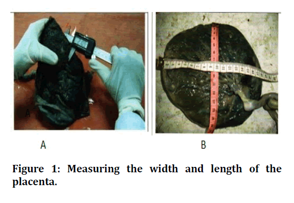
Well-fixed bits of I, II &III trimester placentas were processed histologically and embedded. Paraffin section of 6μm were cut and stained using Haematoxylin and Eosin stain and observed using light microscope and oculomicrometer, the results are tabulated in Table 1 and Figure 1A and 1B.
Table 1: Paraffin section of 6µm were cut and stained using Haematoxylin and Eosin stain and observed using light microscope and oculomicrometer.
| Dimensions | Maximum | Minimum | Average |
|---|---|---|---|
| Length (cm) | 6 | 10 | 7.75 |
| Breadth (cm) | 5 | 12.5 | 7.2 |
| Thickness (cm) | 0.7 | 0.5 | 0.5 |
| Volume (cc) | 15 | 87.5 | 31.1 |
| Weight (gm) | 108 | 150 | 116.8 |

Figure 1: Measuring the width and length of the placenta.
About 33 placentae were circular in shape and remaining 9 were discoid. Average number of cotyledons present were 24. Insertion of the umbilical cord was eccentric in 35, central, marginal in 6 and velamentous in one. The average length of umbilical cord in III and II trimester placentae were 31.7 and 21.5 cm, respectively. The average diameter of umbilical cord in III and II trimester placentae were 1.3 and 0.5 cm, respectively. In term pregnancies, maximum weight of the baby was 3.7 kg and minimum were 2.2 kg with an average of 2.8 kg. In preterm pregnancies, baby weight varied from 1.9 kg to 2.3 kg with an average of 2.1 kg. The histological features reveals that the intervillous space is the widest in I trimester and it is filled with maternal blood. Features of I trimester gradually reduces and features of II trimester emerges. Average number of villi in II trimester placenta in this study is 38, with average size being 500 μm. About 6 capillaries were present per field. Average number of villi in III trimester placenta in this study is 55. Tabulated in Table 2.
Table 2: Histologic features of placenta.
| Features | I Trimester | II Trimester | III Trimester |
|---|---|---|---|
| No of villi/field (l Ox) | 15 | 38 | 55 |
| Size of villi (µm) | 200 | 500 | 800 |
| No of layers in villi | 2 | 2 | 1 |
| No of capillaries | 2 | 6 | 10 |
| Intervillous space | Wider | Wide | Narrow |
| Anchoring villi | Absent | Present | Present |
| Stem villi | Absent | Present | Present |
| Floating villi | Absent | Absent | Present |
| Placental septae | Absent | Absent | Present |
| Nucleated RBC' s | Present | Absent | Absent |
| Endometrial glands | Present | Absent | Absent |
Discussion
From the 51 placentae collected, 27 were from term and 3 from pre-term pregnancies. Remaining 21 included placentae from post-abortion and medical termination of pregnancies of which 9 were from first trimester and 12 were from second trimester. Morphological analysis of placentae of II and III trimesters and histological analysis of the placentae of all the three trimesters were concordant with those reported in the literature [4]. An attempt has been made to assess the placental volume antenatally using two-dimensional ultrasonography by applying volumetric calculations. Placental volume calculated by this method also correlated well with baby weight. Therefore, though it is known that prenatal evaluation of placental volumes using ultrasound imaging has the potential to decrease the number of unexpected fetal demises as recognized by Jauniaux et al. [5] accurate assessment of placental volume is not widely available. In this study also, estimated placental volume (EPV) calculated by ultrasound correlated well with baby weight. Previous studies reported the value of 3D ultrasound in measuring placental volume. He made a longitudinal case-controlled prospective study in 150 pregnant women and concluded that 3D ultrasound measurement of placental volume correlated well with placental weight and birth weight of the baby. This trend will lead to a better understanding of causes of poor pregnancy outcomes, which in tum will lead to better diagnostic and therapeutic approaches to complicated pregnancies [6-10].Conclusion
Placental volume can be very well considered as a surrogate marker of fetal weight. Prenatal evaluation of placental volumes using ultrasound imaging has the potential to decrease the number of unexpected fetal demises.
Today, only a few specialized centres for placental examination exist m India. As the cost of processing and examining placentas decreases, more placentas delivered will be able to be examined by appropriately trained physicians. This trend will lead to a better understanding of causes of poor pregnancy outcomes, which in tum will lead to better diagnostic and therapeutic approaches to complicated pregnancies.
Funding
No funding sources.
Ethical Approval
The study was approved by the Institutional Ethics Committee.
Conflict of Interest
The authors declare no conflict of interest.
Acknowledgement
The encouragement and support from Bharath Institute of Higher Education and Research, Chennai, is gratefully acknowledged. For provided the laboratory facilities to carry out the research work.
References
- Allan C Barnes. Intra-uterine development. Eur J Obstet Gynecol Reprod Biol 1968; 6:325-31.
- Aplin JD. The cell biological basis of human implantation. Baillieres Best Pract Res Clin Obstet Gynaecol 2000; 14:757-64.
- Benirschke K, Kaufmann P, Baergen R. Pathology of the human placenta. 5th Edn. New York: Springer, 2006.
- Bouw GM, Stolte LAM, Baak JPA, et al. Quantitative morphology of the placenta. Standardization of sampling. Eur J Obstet Gynecol Reprod Biol 1976; 6:325-31.
- Jauniaux E, Ramsay B, Campbell S. Ultrasonographic investigation of placental morphologic characteristics and size during the second trimester of pregnancy. Am J Obstet Gynecol 1994; 170:130-137.
- Kaufmann P, Sen DK, Schweikhart G. Classification of human placental villi. Histology. Cell Tissue Res 1979; 200:409-23.
- Moore KL, Persaud TVN. The developing Human-clinically oriented embryology, Elsevier 2008; 134.
- Khong TY, Chambers HM. Alternative method of sampling placentas for the assessment of uteroplacental vasculature. J Clin Pathol 1992; 45:925-7.
- Kliman HJ. Behind every healthy baby is a healthy placenta. In: Berman MR, ed. Parenthood Lost. Westport CT: Bergin & Garvey 2001; 130-131.
- Kucuk M, Doymaz F. Placental weight and placental weight-to-birth weight ratio are increased in diet-and exercise-treated gestational diabetes mellitus subjects but not in subjects with one abnormal value on 100-g oral glucose tolerance test. J Diabetes Complicat 2009; 23:25-31.
Author Info
Sree Balaji Medical College & Hospital Affiliated to Bharath Institute of Higher Education and Research, Chennai, Tamil Nadu, IndiaCitation: Arathi MS, A Study of Morphology and Histological Features of Various Trimesters of Placenta and to Correlate with Ultrasound, J Res Med Dent Sci, 2021, 9(7): 287-288
Received: 07-Jul-2021 Accepted: 22-Jul-2021
