Research - (2021) Volume 9, Issue 5
A Stereomicroscopic Evaluation of Four Endodontic Sealers Penetration into Artificial Lateral Canals Using Gutta-Percha Single Cone Obturation Technique
Omar Jihad Banawi* and Raghad Abdulrazzaq Alhashimi
*Correspondence: Omar Jihad Banawi, Department of Conservative Dentistry College of Dentistry, University of Baghdad, Iraq, Email:
Abstract
Background: Three-dimension obturation of whole root canals system complexity is so important step in root canal filling to isolate the remnant microorganisms from space and neutrinos and consequently is the success of such treatment
Aim of the study: Evaluation the filling capacity of artificial lateral canals after main canal single cone obturation with the following endodontic sealers:
TotalFill® BC, Gutta Flow bioseal, MTA fill apex and AH- plus forty single-rooted extracted lower premolars were instrumented up to size X2 (protaper Next, Dentsply) then generate three lateral canals in each mesial and distal aspects then randomly divided into four gropes (n=10) each group was obturated with single cone gutta-percha with deferent sealers G1: calcium silicate based sealer TotalFill® BC Sealer™(FKG), G2: ROEKO GuttaFlow® Bioseal (COLTEN), G3: MTA Fillapex(ANGELUS) and G4: AH Plus® (DENTSPLY)
The teeth will be demineralizing with week acid to evaluate the penetration of sealers into artificial lateral canals by stereomicroscope the significance of the difference of different means (quantitative data) were tested using Studentst-test for the difference between each two-independent means.
Result: For all ALCs of roots, the GUTTAFLOW BIOSEAL sealer had the highest mean penetration (94.83 ± 6.29) which was significantly higher than the penetration of AH-PLUS (p-value0.030), MTA-FILLAPEX (p-value 0.0001) and Totallfill BC (p-value0.001).
GUTTAFLOW BIOSEAL sealer In the coronal third had the highest mean penetration which was significantly higher than the penetration of Totallfill BC (p-value 0.049) and MTA-FILLAPEX (p-value 0.002) and not significant with AHPLUS (p-value 0.227).in the middle third also significantly higher than the penetration of Totallfill BC (p-value 0.017) and MTA-FILLAPEX (p-value 0.007) and not significant with AH-PLUS (p-value 0.278).the same in apical third which was significantly higher than the penetration of Totallfill BC (p-value 0.0001) and MTA-FILLAPEX (p-value 0.003) and not significant with AH-PLUS (p-value 0.054).
Keywords
Artificial lateral canals ALCs, Gutta flow® bioseal, Demineralization process
Introduction
The Root canal system is morphologically complicated because it contains many ramifications, apical delta, isthmuses, accessory and/or lateral canals, that makes the accurate cleaning and shaping of the affected tooth are difficult, and subsequently, the appropriate sealing by endodontic material [1,2]. The major Endodontic treatment goal is apical periodontitis prevention and treatment, and the way to achieve such treatment is the disinfection and filling the root canal system thoroughly [3]. Due to these challenges, it is paramount important to concentrate on the Root canal–filling Procedures via adequate filling of the root canal system including both main and lateral canals especially just like the apical foramen, lateral canals can create links between the main root canal and surrounding periodontal ligament [4]. The endodontic literature shows that the rate of Lateral canals are at around 27.4-45% of all teeth and most of them in the apical third of the roots are found [2,4]. Some authors concluded that no relationship between the not well filed lateral canals and the periodontal ligament inflammation [5], others demonstrated poorly filed lateral canals pathogenicity after remission of periradicular lesions in relation with the well filling of lateral canals [4,6,7].
Unremoved bacteria in root canal treated teeth may be in instrumented parts of the root canal system like accessory canals [6]. therefore, root canal system three-dimensional obturation becomes extremely important, to prevent reinfection [8] and the microbes can be isolated in inac¬cessible areas, to isolate it from nutrition [9].
Gutta-percha with a root canal sealer is the most common material used in root canal system obturation. The sealer work is to fill the minor irregularities and acts as a luting agent between the gutta-percha and canal wall. An endodontic sealer should have many of characteristic to be considered suitable. the property of Flowability is very important as it reveals its ability to infiltrate small irregularities, accessory canals and dentinal tubules of the root canal system therefore the chosen of the sealers.
TotalFill® BC which was pre-mixed bioceramic sealer BC sealer has good flow properties [10]. The gutta-percha used as plugger to allow hydraulic movement of the sealer into the irregularities of the root canal and accessory canals [11]. Secondly, GuttaFlow Bioseal was new hybrid sealer that is a mixture of polydimethylsiloxane and gutta-percha (GP) powder with calcium silicate particles added, which has Thixotropic property enable sealer to penetrate narrow canals and the tiny ramifications [12].
Third sealer MTA fill apex which was a resinbased sealer, since its composition is primarily resin [13], having suitable flow property [14]. Fourth sealer was AH Plus (AHP) which is a gold standard epoxy resin-based sealer that has been used in literatures due to its excellent physicochemical properties [15].
Materials and Methods
Freshly extracted forty mandibular second premolar were collected from deferent dental centers the criteria for selection of the teeth were free of caries, straight non-classified canal system, absence of lateral canals normal not open apex this done by a radiographic picture taken from the buccolingual direction and by direct inspection of the teeth through Univet 5X magnifying loupes. To standardize the length of the roots the crowns of the teeth were removed with a low- speed diamond disc under running water, and a standard length of 12mm was achieved for every root.
The working length was established by subtracting 0.5mm from root length. Size 10 K-file was used to determine the initial size of the canal. shaping of the canals was done by ProTaper next system (Dentsply Maillefer). A contra-angle handpiece was used with an electric motor fixed on dental surveyor X1 with a tip size of 17mm used after creation glide path then X2 file with 25mm was used as the final file to create 0.06 constant root canal tapering. follow each rotary instrument, the canals were irrigated with 1 mL 5.25% NaOCl by inserting a 27-G needle 3 mm short of the working length without any binding of the needle to the canal wall to allow backflow of irrigation solution easily The smear layer was eliminated by applying 5 mL 17% EDTA for 60 s, finally irrigation with 5 mL 5.25% NaOCl, and generous washing with 5 mL of normal saline. after that three pairs of artificial lateral canals ALCs were prepared in the proximal sides of the roots (mesial and distal) at 2mm (apical third),4mm (middle third) and 6mm (coronal third) using engine reamer (0.1/02)
To conform the patency of ALCs k file size 8 was used also 17% EDTA with ultrasonic activation (passive ultrasonic activation PUA) three-cycle activation was done each one 20 sec to conform ALCs patency finally, the root canal system was washed with normal saline. The shaped roots were randomly divided into four groups (n=10) depending on the type of the sealer used with single cone obturation technique each sample was submerged 9mm in vinyl polysiloxane impression material (putty consistency) that placed inside a plastic tube to mimic the role of the periodontal ligament and confined the sealer while penetrating the ALCs.
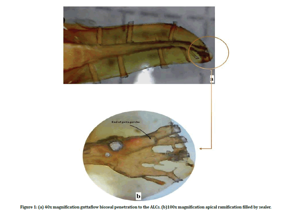
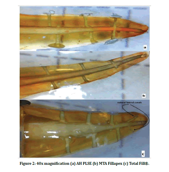
The application of the sealers to the shaped canals applied through lentulo spiral file size 25 twice the time to ensure enough sealer and the end of gutta-percha cone was dipped with sealer and insert it to the WL of the shaped canal, samples were obturated with gutta-percha of the corresponding size and taper X2 Dentsply. The samples were storage for 7 days at 37°C and 100% relative humidity in an incubator. Then the roots were undergoing demineralization process with weak acid and buffering solution which was 7% formic acid; 3% hydrochloric acid; and 8% sodium citrate in aqueous solution with continuous agitation for 14 days the solution was refreshing every 3 days then the samples were storage one day in 99% acetic acid. After that, all samples were rinsed with tap water for 2 hours and dehydrated with 25, 50, 70, 90, 95 and 100% ethanol (30 min passage each) finally the samples were cleared and diaphanized with methyl-salicylate. All ALCs were analyzed with a stereomicroscope at 40X magnification see Figures 1 and 2.

Figure 1: (a) 40x magnification guttaflow bioseal penetration to the ALCs. (b)100x magnification apical ramification filled by sealer.s

Figure 2: 40x magnification (a) AH PLSE (b) MTA Fillapex (c) Total Fill®.
With image processing program image J, the penetration ALCs was measured linearly, using a scale in millimeters. The values that obtained were divide by the total length of the lateral canal and multiply by 100 to obtain the percentage of obturation after the use of each sealer.
Results
Data were presented in simple measures of frequency, percentage, mean, standard deviation, and range (minimum-maximum values). The significance of the difference of different means (quantitative data) were tested using Students-ttest for the difference between two independent means Table 1.
Table 1: Sealer type penetration Comparison in apical, middle, coronal and all 3rd and the average of penetration.
| Endodontic sealer type | Average% Penetration into Artificial Lateral Canal of 3rd | P value compared to | ||
|---|---|---|---|---|
| Apical | ||||
| Average | TOT | GUT | MTA | |
| AH PLUS | 79.17 ± 24.99 (29.4-100) | 0.015* | 0.054 | 0.129 |
| Totalfill BC | 38.20 ± 41.22 (0-100) | 0.0001* | 0.252 | |
| GUTTAFLOW BIOSEAL | 96.32 ± 8.13 (74.1-100) | 0.003* | ||
| MTAFILLAPEX | 58.09 ± 33.58 (8.65-100) | |||
| Middle | ||||
| Average | ||||
| AH PLUS | 86.65 ± 17.53 (50-100) | 0.069 | 0.278 | 0.038* |
| Totalfill BC | 61.82 ± 36.61 (15.53-100) | 0.017* | 0.998 | |
| GUTTAFLOW BIOSEAL | 95.00 ± 15.81 (50-100) | 0.007* | ||
| MTAFILLAPEX | 61.78 ± 30.53 (0-100) | |||
| Coronal | ||||
| Average | ||||
| AH PLUS | 82.90 ± 23.47 (33-100) | 0.29 | 0.227 | 0.061 |
| Totalfill BC | 68.09 ± 35.97 (0-100) | 0.049* | 0.575 | |
| GUTTAFLOW BIOSEAL | 93.18 ± 11.14 (72.75-100) | 0.002* | ||
| MTAFILLAPEX | 59.89 ± 27.77 (20.3-100) | |||
| All ALCs | ||||
| Average | ||||
| AH PLUS | 82.90 ± 14.67 (61-100) | 0.026* | 0.030* | 0.011* |
| Totalfill BC | 56.04 ± 31.75 (15.48-100) | 0.001* | 0.75 | |
| GUTTAFLOW BIOSEAL | 94.83 ± 6.29 (81.37-100) | 0.0001* | ||
| MTAFILLAPEX | 59.92 ± 20.83 (20.58-86.6) | |||
*Significant difference between two independent means using Students-t-test at 0.05 level.
Data were presented as Mean ± SD (Range)
TOT (Totalfill BC), GUT (GUTTAFLOW BIOSEAL), MTA(MTAFILLAPEX), ALCs (Artificial lateral canals)
In all ALCs of roots, the GUTTAFLOW BIOSEAL sealer had the highest mean penetration (94.83 ± 6.29) which was significantly higher than the penetration of AH-PLUS (82.90 ± 14.67), MTA-FILLAPEX (59.92 ± 20.83) and Totallfill BC (56.04 ± 31.75). Totallfill BC had the lowest penetration mean (56.04 ± 31.75) which was significantly not different from MTA-FILLAPEX (59.92 ± 20.83). The mean penetration using AH-PLUS was (82.90 ± 14.67) which was significantly more than MTA-FILLAPEX (59.92 ± 20.83) and Totallfill BC (56.04 ± 31.75) but significantly less than GUTTAFLOW BIOSEAL. The mean penetration using MTA-FILLAPEX was (59.92 ± 20.83) which was significantly not different from Totallfill BC (56.04 ± 31.75) but it significantly less than GUTTAFLOW BIOSEAL and AH-PLUS as mentioned above in the apical area The GUTTAFLOW BIOSEAL sealer had the highest mean penetration (96.32 ± 8.13), which was significantly higher than the penetration of MTA-FILLAPEX (58.09 ± 33.58) and Totallfill BC (38.20 ± 41.22) and not significant with AHPLUS (79.17 ± 24.99).
Totallfill BC had the lowest penetration mean (38.20 ± 41.22) which was significantly not different from MTA-FILLAPEX (58.09 ± 33.58). The mean penetration using AH-PLUS was (79.17 ± 24.99) which is not significant with GUTTAFLOW BIOSEAL (96.32 ± 8.13) and MTAFILLAPEX (58.09 ± 33.58) but it significantly more than Totallfill BC (38.20 ± 41.22). The mean penetration using MTA-FILLAPEX was (58.09 ± 33.58) which is not significant with AHPLUS (79.17 ± 24.99) and Totallfill BC (38.20 ± 41.22) but it significantly less than GUTTAFLOW BIOSEAL (96.32 ± 8.13) the middle area the GUTTAFLOW BIOSEAL sealer had the highest mean penetration (95.00 ± 15.81) which was significantly higher than the penetration of Totallfill BC (61.82 ± 36.61) and MTA-FILLAPEX (61.78 ± 30.53) and not significant with AH-PLUS (86.65 ± 17.53). The MTA-FILLAPEX had the lowest penetration mean (61.78 ± 30.53) which was significantly not different from Totallfill BC (61.82 ± 36.61).
The mean penetration using AH-PLUS was (86.65 ± 17.53) which is not significant with GUTTAFLOW BIOSEAL (95.00 ± 15.81) and Totallfill BC (61.82 ± 36.61), but it significantly more than MTA-FILLAPEX (61.78 ± 30.53). The mean penetration using Totallfill BC was (61.82 ± 36.61) which is not significant with AH-PLUS (86.65 ± 17.53) and MTA-FILLAPEX (61.78 ± 30.53) but it significantly less than GUTTAFLOW BIOSEAL as mentioned above. While In the coronal third of the roots, the GUTTAFLOW BIOSEAL sealer had the highest mean penetration (93.18 ± 11.14) which was significantly higher than the penetration of Totallfill BC (68.09 ± 35.97) and MTA-FILLAPEX (59.89 ± 27.77) and not significant with AH-PLUS (82.90 ± 23.47).
The MTA-FILLAPEX had the lowest penetration mean (59.89 ± 27.77) which was significantly not different from AH-PLUS (82.90 ± 23.47) and Totallfill BC (68.09 ± 35.97). The mean penetration using AH-PLUS was (82.90 ± 23.47) which was not significant with GUTTAFLOW BIOSEAL (93.18 ± 11.14), Totallfill BC (68.09 ± 35.97) and MTA-FILLAPEX (59.89 ± 27.77). The mean penetration using Totallfill BC was (68.09 ± 35.97) which was not significant with AH-PLUS (82.90 ± 23.47) and MTA-FILLAPEX (59.89 ± 27.77) but it significantly less than GUTTAFLOW BIOSEAL as mentioned above.
Discussion
The overwhelming success of endodontic treatment is dependent upon indispensable factors including, three dimensions and adequate obturation with thorough sealing of the root canal system which prevent percolation of microbes into the root canal's space, providing a favorable biological condition for healing [16]. The endodontic literature claims that the adequate seal of lateral and accessory canals is a challenge to fill it throughout root canal obturation. A non-filled lateral and accessory canals make a two-way passage for microbes and tissue products between the root canal system and surrounding periodontium, so the lateral canals are considered a constant challenge to endodontists [4].
The chemical and physical properties of the sealers were affected on its penetration to the root canal irregularities, ramifications, accessory and lateral Canals [17] as well the irrigants [18]. The present study used freshly human extracted teeth instead of epoxy resin blocks because of its ease of making narrow ramifications and getting a more accurate simulation of the clinical situation as the surface texture and condition could influence the flow properties of guttapercha and sealer [19] engine reamer size 0.1mm with 0.2 tapers to produce ALCs with 0.2 external taper, which is so small and almost undetectable external taper compared to the most cylindrical shape canals. The single-cone obturation technique was used to assess only the penetration capability of the sealers because the heated gutta-percha that used in warm techniques not only changed the flow characteristic of sealers due to the elevated temperatures, nevertheless enabled easier access of the alpha phase gutta percha to the lateral canals, may generate falsepositive results.
GuttaFlow bioseal showed the highest mean penetration in the ALCs for all three aspects of the roots (apical, middle and coronal) and significantly higher than TotalFill® BC in agreement with Zahid and Ghareeb, 2019 that showed in the middle portion there was a statistically significant difference among sealers with the deepest penetration was found in GuttaFlow bioseal, shallowest penetration was for Endosequence BC [20]. Also, when compare the penetration capacity of the calcium silicate base sealer (iRoot SP) to the artificial lateral canals between a single cone and continuous wave of condensation (thermoplasticized gutta-percha), the significance toward the hot technique [21]. The single cone obturation technique depends on the sealer to obturate and fill the irregularities [22], and Wu et al. [23] found the sealer volume required more in single cone obturation than other techniques [23].
The highest penetration of the GuttaFlow bioseal and the lowest for TotalFill® BC may be due to the delivery technique of the sealers to the canal, lentulo spiral file used for sealers delivery to the shaped root canal, thixotropic behaver of the sealer explain the thick consistency of the GuttaFlow bioseal and more volume of sealer attached to the file before file insertion, then when the file was rotated in a spiral mode and has an action that it pushes the sealer centrifugally as well as the high volume of sealer, the force result from lentulo file lead to decreased in sealer viscosity, increased its flowability and enhanced the penetration capacity to the ALCs.
In the science of rheology, the behaviour of the fluid classified as Newtonian, pseudoplastic and dilatant. In the Newtonian, the force does not affect the viscosity like in water. Otherwise, the viscosity increases when force increased in the dilatant. And in the pseudoplastic, the viscosity will decrease when the force is applied [24].
Thixotropic behaviour is the same as pseudoplastic, but it's time-dependent [24], which mean it takes time to return to its original viscous state after force cessation [25]. This may explain the high rate of GuttaFlow bioseal penetration to the ALCs because the low viscosity of the sealer that results from lentulo file takes time to return its original state permit sealer to penetrate more ALCs. While the TotalFill® BC has a pseudoplastic behaviour [26] mean the viscosity return to its original state after force cessation, as well as the high viscosity that can be attributed to the presence of calcium silicate.
Also, Soma et al. informed more sealer volume in partially oval-shaped canals than round canals [22]. Since the canals were all round that used in the study mean more volume of sealer required that the lentulo file may not offer this requirement for the TotalFill® BC, especially many practitioners use delivery tip to dispense enough bioceramic sealer [27]. Therefore, the force is receded both by the decrease of the pressure on the walls by taper increased, as well as by the loss of mass of the sealer, resulting in the flowability redaction and incomplete filling of the lateral canals. On the other hand, recent studies showed good penetration of the bioceramic sealer to the dentinal tubules independent of the obturation techniques [28,29] because the particles size of this sealer less than 1 μm, enable it to penetrate the tubules measuring about 2 to 3.2 μm in diameter [26]. AH PLUS penetration in the coronal was no significant difference with other sealers in agreement with Candeiro et al. 2019 as mentioned did not significantly penetrate the ALCs between AH PLUS and Endosequence BC sealer [26].
Also Almeida etal.,2007 found no significant difference among five sealers penetration of AH Plus, Endomethasone Epiphany Root Canal Sealant, Pulp Canal Sealer (EWT) and Sealapex to the ALCs [30]. But these results disagreed with Al-Azzawi, et al. who found AH Plus penetration depth more statistically significant than calcium silicate-based sealer IRoot SP in both straight and curved canal but in apical part, the results were in agreement with the present study that AH Plus penetration depth more statistically significant than calcium silicate-based sealer [31].
This probably due to the presence of epoxy resin in the composition of AH Plus that might be responsible for the increase of its viscosity MTA-FILLAPEX penetration rate for all ALCs was statistically less significant than AH PLUS, and GuttaFlow bioseal in disagreement with Melo et al. study that mentioned no statistically difference among Endofill, Sealer 26 and MTA Fillapex and in the coronal third MTA Fillapex showed a significantly greater penetration than Sealer [32], but the ALCs diameter was 0.15 mm and the obturation technique was lateral condensation could influence on the penetration of the sealers in addition to that the investigation method of filled ALCs was done by radiograph which can give falsepositive results [21] which they found 11.5% of ALCs rated with an acceptable score by digital radiograph had an unacceptable score when the using of stereomicroscope after demineralization.
Conclusion
With the limitation of this study and according to the proposed methodology and based on the finding of this in vitro study, the following conclusions could be down:
For all ALCs GuttaFlow bioseal had a higher penetration rate statically more than all other sealers. While AH PLSE, TotalFill®BC and MTA Fillapex were statically not different.
✓ In the apical third of the roots, GuttaFlow bioseal and AH PLUSE had adequate penetration to ALCs, statistically not different. While MTA Fillapex and TotalFill®BC had a lower capacity, which statistically not different from each other.
✓ In the middle third, GuttaFlow bioseal had a higher penetration rate statically more than TotalFill®BC and MTA Fillapex with no differences with AH PLSE.AH PLSE and TotalFill®BC statically not deferent, while MTA Fillapex had the lowest penetration rate statically less than GuttaFlow bioseal and AH PLSE.
✓ In coronal third GuttaFlow had a higher penetration rate statically more than TotalFill®BC and MTA Fillapex with no differences with AH PLSE.There was no statically difference among AH PLSE, TotalFill®BC and MTA Fillapex
✓ Single cone obturation with GuttaFlow bioseal sealer is a reliable technique that can fill the lateral canals in the apical, middle and coronal aspects of the roots.
✓ According to the penetration rate to the ALCs GuttaFlow bioseal and AH PLSE had suitable flow property more than MTA Fillapex and TotalFill®BC.
References
- Peters OA, Peters CI, Schonenberger K, et al. Protaper rotary root canal preparation: Assessment of torque and force in relation to canal anatomy. Int Endod J 2003; 36:93-99.
- Venturi M, Di Lenarda R, Prati C, et al. An in vitro model to investigate filling of lateral canals. J Endod 2005; 31:877-881.
- Nguyen T. Obturation of the root canal system. Pathways Pulp 1994; 6:219-271.
- De Deus Q. Frequency, location, and direction of the lateral, secondary, and accessory canals. J Endod 1975; 1:361-366.
- Barthel CR, Zimmer S, Trope M. Relationship of radiologic and histologic signs of inflammation in human root-filled teeth. J Endod 2004; 30:75-79.
- Weine F. The enigma of the lateral canal. Dent Clin North Am 1984; 28:833-852.
- Ricucci D, Loghin S, Siqueira JF. Exuberant biofilm infection in a lateral canal as the cause of short-term endodontic treatment failure: Report of a case. J Endod 2013; 39:712-718.
- Pinheiro E, Gomes B, Ferraz C, et al. Microorganisms from canals of root-filled teeth with periapical lesions. Int Endod J 2003; 36:1-11.
- Vivacqua‐Gomes N, Gurgel‐Filho E, Gomes B, et al. Recovery of Enterococcus faecalis after single‐or multiple‐visit root canal treatments carried out in infected teeth ex vivo. Int Endod J 2005; 38:697-704.
- Almeida LHS, Moraes RR, Morgental RD, et al. Are premixed calcium silicate–based endodontic sealers comparable to conventional materials? A systematic review of in vitro studies. J Endod 2017; 43:527-535.
- Trope M, Bunes A, Debelian G. Root filling materials and techniques: bioceramics a new hope? Endod Topics 2015; 32:86-96.
- Zhong X, Shen Y, Ma J, et al. Quality of root filling after obturation with gutta-percha and 3 different sealers of minimally instrumented root canals of the maxillary first molar. J Endod 2019.
- Komabayashi T, Colmenar D, Cvach N, et al. Comprehensive review of current endodontic sealers. Dent Materials J 2020; 39:703-720.
- Vitti RP, Prati C, Silva EJNL, et al. Physical properties of MTA Fillapex sealer. J Endod 2013; 39:915-918.
- Tanomaru-Filho M, Torres FFE, Chávez-Andrade GM, et al. Physicochemical properties and volumetric change of silicone/bioactive glass and calcium silicate–based endodontic sealers. J Endod 2017; 43:2097-2101.
- Al-Hashimi RA. A micro computed tomography assessment of new carrier-based root canal fillings. J Baghdad College Dent 2015; 325:1-4.
- Venturi M. An ex vivo evaluation of a gutta-percha filling technique when used with two endodontic sealers: Analysis of the filling of main and lateral canals. J Endod 2008; 34:1105-1110.
- Guimarães BM, Amoroso-Silva PA, Alcalde MP, et al. Influence of ultrasonic activation of 4 root canal sealers on the filling quality. J Endod 2014; 40:964-968.
- Reader CM, Himel VT, Germain LP, et al. Effect of three obturation techniques on the filling of lateral canals and the main canal. J Endod 1993; 19:404-408.
- Zahid HM, Ghareeb NH. Evaluation of filling ability of Guttaflow Bioseal sealer to the simulated lateral canal by scanning electron microscope. Erbil Dent J 2019; 2:218-228.
- Fernández R, Restrepo J, Aristizábal D, et al. Evaluation of the filling ability of artificial lateral canals using calcium silicate‐based and epoxy resin‐based endodontic sealers and two gutta‐percha filling techniques. Int Endod J 2016; 49:365-373.
- Somma F, Cretella G, Carotenuto M, et al. Quality of thermoplasticized and single point root fillings assessed by micro‐computed tomography. Int Endod J 2011; 44:362-369.
- Wu MK, Bud MG, Wesselink PR. The quality of single cone and laterally compacted gutta-percha fillings in small and curved root canals as evidenced by bidirectional radiographs and fluid transport measurements. Oral Surg Oral Med Oral Pathol Oral Radiol Endod 2009; 108:946-951.
- Sakaguchi RL, Powers JM. Craig's restorative dental materials-e-book: Elsevier Health Sciences 2012.
- Morrison I. Dispersions. Kirk‐othmer encyclopedia of chemical technology 2000.
- Candeiro GTDM, Lavor AB, Lima ITDF, et al. Penetration of bioceramic and epoxy-resin endodontic cements into lateral canals. Br Oral Res 2019; 33.
- Koch K, Brave D, Nasseh AA. A review of bioceramic technology in endodontics. CE Article 2012; 4:6-12.
- Coronas VS, Villa N, Nascimento ALD, et al. Dentinal tubule penetration of a calcium silicate-based root canal sealer using a specific calcium fluorophore. Br Dent J 2020; 31:109-115.
- Yang R, Tian J, Huang X, et al. A comparative study of dentinal tubule penetration and the retreat ability of endo sequence BC sealer HiFlow, iRoot SP, and AH Plus with different obturation techniques. Clin Oral Investigations 2021; 1-11.
- Almeida J, Gomes B, Ferraz C, et al. Filling of artificial lateral canals and microleakage and flow of five endodontic sealers. Int Endod J 2007; 40:692-699.
- Al-Azzawi AJ. Comparison of three endodontic sealers penetration into simulated lateral canals. J Genetic Environment Resources Conservation 2014; 2:349-355.
- Melo TA, Nunes DP, Al-Alam FD, et al. Filling analysis of artificial lateral canals after main canal obturation through three different endodontic sealers. RSBO 2014; 11:377-382.
Author Info
Omar Jihad Banawi* and Raghad Abdulrazzaq Alhashimi
Department of Conservative Dentistry College of Dentistry, University of Baghdad, IraqCitation: Omar Jihad Banawi, Raghad Abdulrazzaq Alhashimi, A Stereomicroscopic Evaluation of Four Endodontic Sealers Penetration into Artificial Lateral Canals Using Gutta-Percha Single Cone Obturation Technique, J Res Med Dent Sci, 2021, 9 (5):57-64.
Received: 27-Apr-2021 Accepted: 13-May-2021 Published: 31-May-2021
