Case Report - (2022) Volume 10, Issue 9
A Rare Case of Thoracic Disc Prolapse at D1-D2 Level Presented with Right Shoulder Pain Managed by Disc Discectomy
Akilan SS*, Ganesh MT and Vasanth kumar
*Correspondence: Akilan SS, Department of Orthopaedics, Sree Balaji Medical College and Hospital, Bharath Institute of Higher Education and Research, Chennai, Tamil Nadu, India, Email:
Abstract
Aim: The main aim of the study is to prospectively analyse the functional outcome of thoracic disc prolapse at D1-D2 level presented with right shoulder pain managed by disc discectomy.
Methods: A prospective analysis of 10 patients (6 men and 4 women, with mean age 35 years) with anatomic classification thoracic disc prolapse at D1-D2 level presented with right shoulder pain managed by disc discectomy. Procedure between July 2019 and July 2021 was performed. The outcomes were studied based on Neurological Assessment Score (NAS) and Visual Analog Score (VAS).
Results: The mean NAS and VAS at the interval of 3 weeks, 5 weeks, 7 weeks, 6 months, 1 year shows excellent improvement. The mean VAS score is 8.8 and NAS is excellent.
Conclusion: In the present study, 10 patients with thoracic disc prolapse at D1-D2 level presented with right shoulder pain managed by disc discectomy. Management of thoracic disc prolapse at D1-D2 level presented with right shoulder pain managed by disc discectomy allows for early rehabilitation of the patient and has excellent functional outcome with less incidence of complications. Hence, we strongly recommend considering it in the management of thoracic disc prolapse at D1-D2 level.
Keywords: Thoracic disc prolapse, D1-D2 Level, Discectomy, Shoulder pain, Neurological Assessment Score (NAS) and Visual Analog Score (VAS)
Introduction
Disc prolapse in upper thoracic spine is extremely rare clinical condition. Disc prolapse are caused by aging, degeneration of the disc (disc disease) [1]. Disc disease may result from tiny tears or cracks in the outer capsule of the disc called annulus. Magnetic resonance imaging is the most important and useful imaging method to show thoracic disc prolapse. A 35 year old male presented with complaints of right sided shoulder pain for 3 months and right sided scapular pain for 3 months. Patient was heavy labourer by occupation [2]. There was no history of any injury. Initially radiculopathy symptoms were not there and hence the shoulder pathology was suspected and managed with analgesics, with no improvement in symptoms. As the symptoms progressed further along with right arm pain, cervical radiculopathy was suspected and appropriate investigations x-ray and MRI cervical spine with whole spine screening were taken. Surprisingly no cervical disc prolapse was noted. However, in upper thoracic MRI there was an extruded disc prolapse at D1-D2 level compressing the right t1 nerve root. The clinical picture correlated with MRI findings and hence the diagnosis of D1-D2 disc prolapse was made. A trial of conservative management showed no clinical improvement. Hence surgical management was planned and D1-D2 discectomy was done through posterior approach. There was excellent improvement in symptoms immediately after the surgery [3].
From this it is evident that cardiothoracic or upper thoracic disc disease may present with symptoms similar to cervical radiculopathy or shoulder pathology. Through clinical examination and proper MRI is very important in diagnosing such uncommon presentation [4].
Aim and objective: The main purpose of the study is to prospectively analyse the functional outcome of thoracic disc prolapse at D1-D2 level presented with right shoulder pain managed by disc discectomy.
Methods: There are 6 patients with an age group of 25 to 40 years of age. Thoracic disc prolapse was classified on anatomic classification and treatment is then processed. Out of these, 2 Patients were thoracic disc prolapse presented with shoulder pain. Patient was treated with Disc Discectomy [5].
Case Presentation
A prospective analysis of 10 patients (6 men and 4 women, with mean age 35 years) with Anatomic classification thoracic disc prolapse at D1-D2 level presented with right shoulder pain managed by disc discectomy. Procedure between July 2019 and July 2021 was performed. The outcomes were studied based on Neurological Assessment Score (NAS) and Visual Analog Score (VAS).
Informed consent and written consent were taken from all patients. Surgery was done electively after assessment under regional anaesthesia. All cases were taken up for surgery immediately following admission [6].
Methodology
The management method was decided after classifying Thoracic disc prolapse by Anatomic classification. The patients were taken for surgery as early as possible time depending on their co morbidities and skin condition. Thoracic disc prolapse were classified according to Anatomic classification [7].
Pre-operative preparation: patient underwent a pre-operative evaluation including the following parameters: Hb, blood sugar, ECG, Renal function test, X ray Chest in order to get fitness for surgery.
All surgeries were done under C-arm guidance. Thoracic Disc Prolapse was managed by Disc Discectomy.
Follow up period: At 1 week, 3 weeks, 5 weeks, 1 month, 3 months, and 5 months.
Results
The mean NAS and vas at the interval of 1 week, 3 weeks, 5 weeks, 1 month, 3 months and shows excellent improvement. Mean vas score is 8.8 and range of motion is excellent.
The result shows the efficacy of the functional outcome of thoracic disc prolapse at D1-D2 level presented with right shoulder pain managed by disc discectomy.
Discussion
A 35 years old male presented with complaints of right sided shoulder pain for 3 months and right sided scapular pain for 3 months. Patient was heavy labourer by occupation. There was no history of any injury. Initially radiculopathy symptoms were not there and hence the shoulder pathology was suspected and managed with analgesics, with no improvement in symptoms.as the symptoms progressed further along with right arm pain, cervical radiculopathy was suspected and appropriate investigations x-ray and MRI c. spine with whole spine screening were taken. Surprisingly no cervical disc prolapse was noted. However, in upper thoracic MRI there was an extruded disc prolapse at D1-D2 level compressing the right T1 nerve root. The clinical picture correlated with MRI findings and hence the diagnosis of D1-D2 disc prolapse was made. A trial of conservative management showed no clinical improvement. Hence surgical management was planned and D1-D2 discectomy was done through posterior approach. There was excellent improvement in symptoms immediately after the surgery (Figures 1-10).
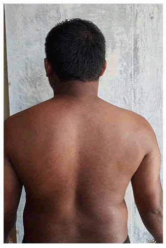
Figure 1: Mild tenderness felt over scapular and Para spinal region and mild kyphosis is seen.
Figure 2: This two finding the radio culopathy symptoms is evident that there might be any Disc pathology mild kyphosis is seen.
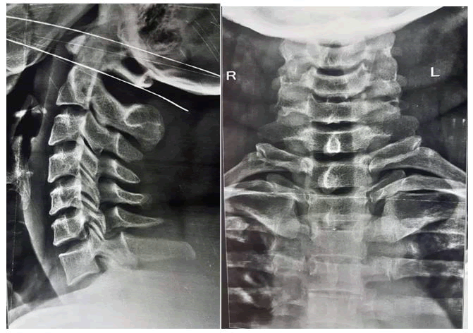
Figure 3: X-ray shows osteophytic changes and disc space is being reduced both in cervical thoracic disc.
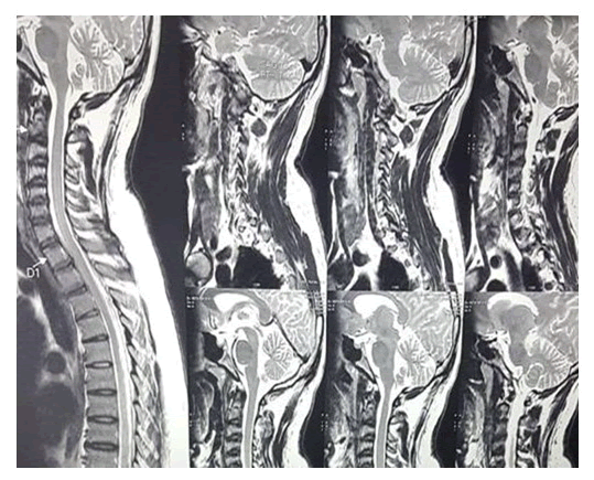
Figure 4: This MRI T2 guided image shows extruded D1-D2 disc prolapse and it compresses D1 root level in sagittal plane.
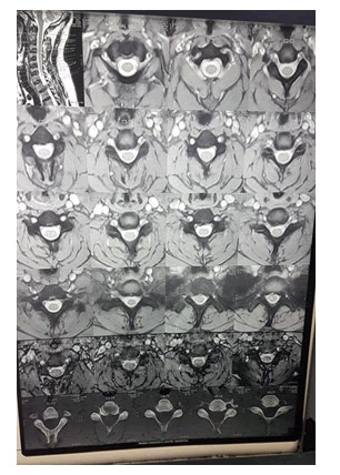
Figure 5: MRI T2 guided image shows D1 root compression in the axial section.
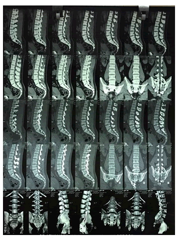
Figure 6: CT image shows osteophytic changes in the cervical and thoracic disc.
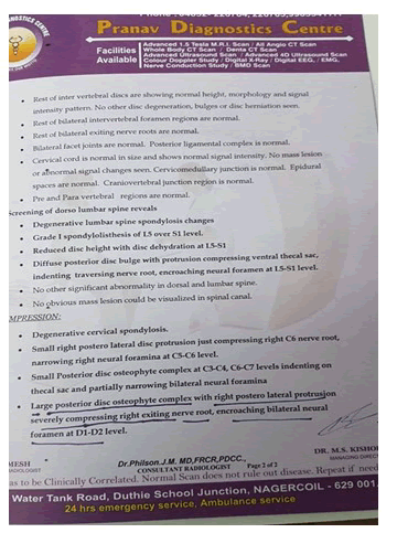
Figure 7: MRI report given by radiologist confirmed D1-D2 prolapse compressing D1 root level.
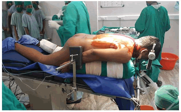
Figure 8: Image shows patient is in prone position for discectomy.
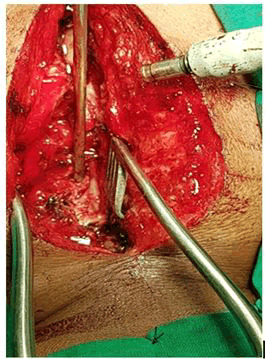
Figure 9: Intraoperativeimage of D1-D2 ruptured fregment being removed.
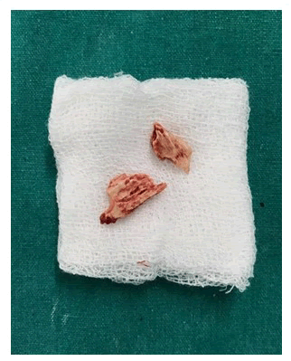
Figure 10: Image shows removed extruded disc fragment.
Radiological evaluation
The symptoms progressed further along with right arm pain, cervical radiculopathy was suspected and appropriate investigations X-RAY and MRI C. Spine with Whole Spine screening were taken. Surprisingly no cervical disc prolapse was noted. However, in upper thoracic MRI there was an extruded disc prolapse at D1-D2 level compressing the right T1 nerve root.
Procedure done: A trial of conservative management showed no clinical improvement.
- surgical management of D1-D2 discectomy done
- Position-prone position
- Approach-posterior approach.
Conclusion
There was excellent improvement in symptoms immediately after the surgery. From this it is evident that cervicothoracic or upper thoracic disc disease may present with symptoms similar to cervical radiculopathy or shoulder pathology. Through clinical examination and proper MRI is very important in diagnosing such uncommon presentation.
An accurate diagnosis and timely surgical intervention may provide the patient the best chance for regression of symptoms and a satisfactory outcome. From this it is evident that cervico thoracic or upper thoracic disc disease may present with symptoms similar to cervical radiculopathy or shoulder pathology. Through clinical examination and proper MRI is very important in diagnosing such uncommon presentation.
References
- Arseni C, Nash F. Thoracic intervertebral disc protrusion: a clinical study. J Neuro surg 1960; 17:418–430.
- Murphey F, Simmons JCH, Brunson B. Surgical treatment of laterally ruptured cervical disc: review of 648 cases, 1939 to 1972. J Neurosurg 1973; 38:679–683.
- Sekhar LN, Jannetta PJ. Thoracic disk herniation: operative approaches and results. Neuro surg 1983; 12:303–305.
- Boriani S, Biagini R, De Lure F, et al. FioreTwo-level thoracic disk herniation. Spine 1994; 21:2461–2466.
- Okada Y, Shimizu K, Ido K, et al. Multiple thoracic disk herniations: case report and review of the literature. Spinal Cord 1997; 35:183–186.
- Arseni C, Nash F. Thoracic intervertebral disc protrusion: a clinical study. J Neurosurg 1960; 17:418–430.
- Svien HJ, Karavitis AL. Multiple protrusions of intervertebral disks in the upper thoracic region: report of case. Proc Staff Meet Mayo Clin 1954; 29:375–378.
Author Info
Akilan SS*, Ganesh MT and Vasanth kumar
Department of Orthopaedics, Sree Balaji Medical College and Hospital, Bharath Institute of Higher Education and Research, Chennai, Tamil Nadu, IndiaCitation: Akilan SS, Ganesh MT, Vasanth kumar, A Rare Case of Thoracic Disc Prolapse at D1-D2 Level Presented with Right Shoulder Pain Managed by Disc Discectomy, J Res Med Dent Sci, 2022, 10 (9): 000-000.
Received: 04-Jul-2022, Manuscript No. JRMDS-22-57347; , Pre QC No. JRMDS-22-57347; Editor assigned: 07-Jul-2022, Pre QC No. JRMDS-22-57347; Reviewed: 21-Jul-2022, QC No. JRMDS-22-57347; Revised: 05-Sep-2022, Manuscript No. JRMDS-22-57347; Published: 12-Sep-2022

