Review Article - (2021) Volume 9, Issue 11
A Paradigm Shift in Designs of Access Cavity Preparations-A Review
Meghna Dugar*, Anuja Ikhar, Pradnya Nikhade, Manoj Chandak, Akshay Jaiswal, Rutuja Rajnekar and Radhika Gupta
*Correspondence: Meghna Dugar, Department of Conservative Dentistry and Endodontics, Sharad Pawar Dental College and Hospital, Datta Meghe Institute of Medical Science (Deemed University), Sawangi(M), Wardha, Maharshtra, India, Email:
Abstract
Access cavity preparations are important to attain longstanding success of endodontically treated teeth. However, this success determining step is sometimes neglected by clinging to the traditional designs which were formulated based on the armamentarium and restorative materials available at that time. It’s the time to shift towards more conservative preparation as nothing can compensate for natural dentin. This review vividly compares traditional approaches and various minimally invasive treatment methods in Endodontics. It focuses on strategies involved in attainment of long lasting of the treatment for successful results. All similar textbooks and articles were looked. Devan quoted once “Our goal should be perpetual preservation of what remains rather than meticulous restoration of what is missing” which could be attained by the shifting the focus from the concept of “extension for prevention” in endodontics to minimal invasion into the tooth structure. Minimally invasive approach requires in-depth knowledge of Root Canal Anatomy, Diagnosis and Decision Making, Preservation of Structural Integrity of Tooth, Alternate Access Designs, Image Guided Endodontic Access, Dynamically Guided Endodontic Access, Microguided Endodontic Access, Modern Bur Designs, Cleaning and Shaping, 3D Irrigation and Disinfection, Root Strengthening and Magnification aids like Loupes and Surgical Operative Microscope.
Keywords
Access designs, Calla lily, Minimally invasive endodontics, Ninja access, Traditional cavityIntroduction
An entry made in a tooth in order to gain ingress into the canal of the root for irrigation as well as obturation is known as an access cavity. A well prepared access cavity allows effective cleaning, shaping as well as obturation of canal of the root. However, on the other hand, an incomplete endodontic access cavity makes the steps of root canal difficult during the treatment from the very beginning i.e to locate the canals to negotiate, to debride, to disinfect, and to obturate the root canals. Hence, lacking a proper access cavity in endodontics, can hamper the outcome of a root canal procedure. There is a significance of minimal access preparation of teeth which primarily aims to retain Pericervical Dentine (PCD) to, reinforces the root canal treated teeth. Hence, for long term biological and functional integrity of root canal treated teeth there is a need to shift from traditional access to more conservative access.
Literature Review
The traditional endodontic access cavity
According to the tooth type, there is a predetermined shape of the traditional endodontic access cavity. As seen in a prepared conventional cavity, the endodontic cavity also consists of an outline which ascertains occlusally to the limit of the cavity. Here, in the cavity, the convenience form is made with respect to removal of structure of tooth done for an access of a straight-line. A design of conventional cavity, an extensionis done for prevention extending the straight-line up till the end of the foramen or primary canal’s curvature. These designs of access cavity were there for a long time period since the diagnostic facilities and imaging were not available. These conventional forms of access designs lead to loss of dentin, thereby weakening the structural construction of the tooth [1]. As it is due to caries, the tooth structure weakens. Therefore, the dentin of the crown or the root should be conserved, and geometry of the root is to be carefully maintained, conserving the strength of tooth treated endodontically. In anterior teeth, initial penetration is approximately in the middle of lingual tooth’s surface, above cingulum and nearly 90 degree to the lingual surface. With this preparation, the access cavity is a rough triangular and ovoid shape. This mimics the pulp chambers anatomy, having pulp horn one mesially and one distally. Although it has been seen that a better straight line access is achieved with an access cavity incisally, the lingual approach is used in order to maintain a good amount of structure of the tooth on the labial surface to maintain the esthetics. After the penetration into the chamber is done, we extend the access cavity in both the dimensions labiolingually and mesiodistally, whereas, in posterior teeth, since the floor of the molars takes a quadrilateral shape, it is clear that a similar shape should be seen in the access cavity. One must keep not forget that when creating the access cavity of the posterior teeth, it should not be triangular, but trapezoidal or quadrangular with corners rounded.
Newer advances in access designs
- Conservative Endodontic Access Cavity
- Ninja Endodontic Access Cavity
- Orifice-Directed Dentin Conservation Access Cavity
- Incisal Access
- Calla Lilly Enamel Preparation
- Image guided endodontic access preparations.
Discussion
Conservative endodontic access cavity (cecs)
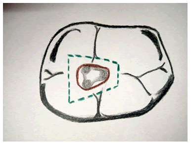
David Clark and Khademi renewed the conventional access cavities as well as developed the conservative or constricted access cavities there by cutting down the remaining structure of tooth and maintaining the tooth’s stability mechanically for long lasting survival as well as function of the teeth treated endodontically [2]. Here, it is seen that the teeth are penetrated centrally at the fossa and then as per need extended inorder to find out canal orifices, so that peri-cervical dentin as well as a pulp chamber floor’s part is preserved, thus aiming to increase the remaining amount of tooth structure (Figure 1). Care should also be taken while instrumentation and using the right type of armamentarium during preparation. The tooth structure’s loss during the endodontic preparations is unable to be replaced with any restorative material completely, therefore, the sole way to maximize the tooth’s strength is dentin conservation. Recently, super flexible diagnostics instruments, magnification microscopes (higher magnification), irrigation kits of high potential, obturation techniques of thermoplasticized kind, as well as most important, CBCT, allowing 3D scanning of the tooth have possibilities of resulting in access cavity of conservative designs. The basic concept of making cavity has changed in this conservative access cavity which makes it more teeth oriented in comparison to the operator oriented cavities concept earlier. Now this cavity depicts a design in which restorative material is taken out before the cutting of the tooth naturally, significantly cutting the dentin after enamel, as well as remove the tooth’s occlusal structure before the remaining cervical dentin. Fracture strengths in mandibular molar prepared according to the traditional endodontic as well as conservative endodontic methods of preparing the cavities [3]. It was noticed that the strength of the teeth, prepared as CEC did not increase with class II cavities in comparison to TEC preparation. The entry into canal of the root is mostly not perpendicular to the surface of the occlusal area. The teeth are entered at central fossa and after which extended till canal orifices are seen preserving chamber roof and peri cervical dentin. Accesses are based on experience / magnification and case difficulty and not based on the outline forms (Figure 1).
Figure 1: A representation of traditional cavity (green dots) and conservative cavity (brown line) in mandibular molar.
Ninja endodontic access cavity (necs)
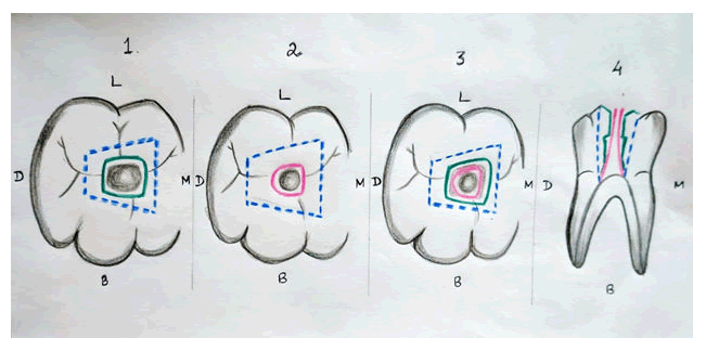
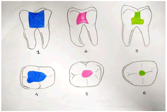
The Ninja access cavity is also called as “PEAC” (point endodontic access cavity) as well as “UEC” (ultraconservative endodontic cavity). ‘Ninja’ outline starts from central fossa and moves towards the canal orifices following an oblique projection (figure 2 and figure 3). It is seen to be in line same as enamel cut which is 90â° or greater, to occlusal area, leaving the root canal orifices tracing from the various visual angulations a lot easier [4]. Gianluca Plotino in a vitro study, compared fracture strength of the restored teeth and root with the conservative cavity, the traditional cavity, or the ninja endodontic access cavity. The results that were seen was that there was a reduce in the fracture probability of the teeth treated endodontically with CEC and NEC .An undistinguishable kind of fracture strength were seen with CEC and NEC, which was greater than teeth with traditional endodontic access cavity. Therefore, we can say that Ninja endodontic access cavity was found to have a better resistance of fracture when compared to conventional access prepared cavity.
Figure 2: 1-4 sketches showing, occlusal view (1-3) and sagittal view (4) of designs of access cavity of lower molars (first). Traditional access cavity (1-4) (blue-dashed line), conservative access cavity (1,3 and 4) (green), and the “ninja” ultraconservative cavity (2-4) (pink).Comparing the 3 kinds of access cavity designs; in no.4 (sagittal view) and in no.3 (occlusal view) respectively. A good portion of pericervical dentin is seen in the sagittal view of conservative access cavity “M”- mesial, “D”- distal, “B” - buccal, “L”- lingual.
Figure 3: 3-dimensional CBCT segmentations and reconstructions of mandibular molars, where preparation is done with different designs of access cavity in the sagittal view and the axial view. Traditional access cavity (blue), conservative access cavity (pink) as well as “ninja” ultraconservative access cavity (green) segments on reconstructions of CBCT.
Dentin conservation and orifice-directed access cavity (truss access cavity)
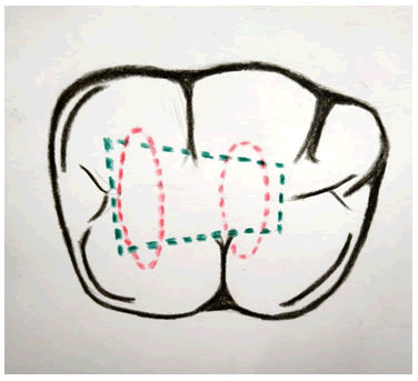
The main motive of the “truss” access cavity design is to leave some amount of dentin between two prepared cavities for preserving the dentin (Figure 4). Different cavities are made in order to approach the canals. Here, we reach the pulp chamber through the crown discontinuities in either caries or a previously done restoration. Hence, it is an approach which is decided by the lesion. It minimizes the restorative necessity of the teeth by taking benefit of the absent hard tissue structures for access. Two separate cavities made preserving the dentin in between the two cavities [5]. The limiting factors of this design of cavity being: inclination of the tooth, complexity of the anatomy, other patient factors etc. For example, in the mandibular molars, we make two different cavities to reach the mesial as well as the distal canals but in the molars, the “mesio-buccal” and the “disto-buccal” canals are reached in a cavity only as well as a complete different cavity for the palatal canal is made. Experts conducted an invitro study of strength of teeth treated endodontically with NECs, TECs or CEC sand saw that both CECs and NECs presented a higher fracture strength than TECs in maxillary as well as mandibular molars and premolars .No particular difference was noticed in mean values of resistance of fracture in NECs and CECs. Traditional cavities leads to a good conservation of the canal’s original anatomy, present while shaping when compared to CECs, especifically at apical portion. Rate of finding out MB2 of traditional (60%), conservative (53.3%) are higher than Ninja (31.6%) cavities statistically. No major differentiation can be made in the fracture resistance of TEC and CEC prepared teeth. However, fracture types are seen to be less serious in case of CEC preparation, Resistance of fracture has been seen higher in cases where cavity is made through the traditional way. The conservation of dentin resulted in an increased fracture resistance in conservative category which is double the resistance of fracture the traditional category. It is clearly seen that in both traditional as well as conservative cavities there are both good and poor results because focusing on too much of conservative cavities can result in improper cleaning and shaping as well as the incapability to get more than the expected number of canals which leads to bad prognosis of the ongoing treatment [6]. Hence, we should strike a balance between both the types of cavities and use a particular design which would result in less failure.
Figure 4: A representation of traditional cavity (green dots) and truss cavity (orange dots) in mandibular molar.
Incisal access
Cavities are prepared on incisal edges than in the cingulum areas in this type of conservative preparation thereby minimising cuspal deformation as well as cusp bending to maintain the dentin bulk and hence restorative needs. Cavities are made the smallest possible thereby maintaining the biologic and mechanical properties during the treatment. Inverse funnelling and blind tunnelling are consequences of the traditional endodontic access which is avoided by a proper access incisally. Blind tunnelling is nothing but gouging observed with highly aggressive round burs. Bucco-lingual gouging (which is not seen in x-rays) occuring in almost every traditional-accessed tooth [7].
The Inverse Funnel; an inverse funnel is made as we move in the access cavity internally. Each time the bur enters the tooth precious peri-cervical dentin is lost.
Calla lily enamel preparation
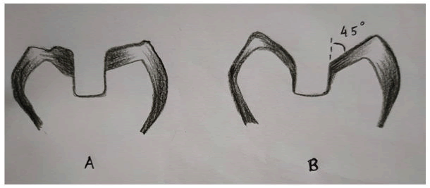
In this type of access cavity preparation, the enamel is cut at 45º in order to engage enamel rods and to provide a favorable C factor. The shape of the preparation resembles a Calla Lily with almost complete involvement of the occlusal surface that aid in resisting the compressive forces (figure 5). Traditional access cavity was compared with Calla Lily enamel preparation and it was seen that unfavourable C factor as well as poor engagement of rods of enamel are present while the old amalgam or composite is removed or in case of the traditional access, which makes 90 degree with the occlusal table. At 45degree, the enamel then is cut in shape of Calla Lily. This preparation allows the involvement of almost entire occlusal surface. The cavosurface should be Calla Lilied if we plan a high bondable substrate like the enamel or porcelain that is etchable as well as the usage of bondable restorative material like the composite resin. If the bondability of the substrate is less, or in cases where it is not possible to establisha bond in between the restorative material and substrate, then the objective should be a butt joint or a 70-90 degree at the cavosurface interface. In cases where multiple visitsare required and where an unbonded temporary restoration is to be placed, a 70-90 degree of the cavosurface is to be maintained until the final visit.
Calla Lily enamel preparation is based on the principle of ICE:
“I”-Infinity edge
“C”-Compression based
“E”-Enamel driven (engage 70% enamel and 30% dentin)
Figure5: Traditional access cavity (parallel-sided) 90° to the occlusal table (A), compared with the Calla Lily access preparation where enamel is cut at 45° (B).
Image-guided endodontic access preparations
It utilizes those images easily accessible to clinicians. Rather than “one common size fitting all”, it ascertains specific location as well as size of access cavity. The purpose is to judiciously preserve dentin and prepare as small an access cavity possible [8]. To customize the kind of access depending on a particular tooth is the ideal action of this system. Image guided endodontic access preparations are of two types mainly;
- CT Dynamic access
- CT/ CBCT guided static 3D templates
Dynamic access: known very commonly as X entry access. It was popularized by Charles M Buchanan. The technique was traditionally used in implantology. The procedure utilizes CBCT volume plan to prepare access by 3D assessment of jaw position and bur position with overhead cameras and software.
Static 3D template: This utilises CBCT images and 3D surface scanners to create virtual images of burs and guide sleeves. A virtual template is designed and printed using 3D printers. Templates are attached to models and access prepared with specially designed burs.
Conclusion
The endodontic access of the traditional type is based on certain principles which go very well with the instruments as well as the restorative materials available at that point of time. However, with the advent of advancements in the materials and instruments there has been a decrease in the amount of tooth structure loss and therefore sticking on to the traditional principles of preparations of access cavity jeopardize the tooth structure. It has also been seen that focusing too much on the minimal technique of cavity preparations can lead to a higher rate of failure in the procedure and could easily outweigh the benefits by producing substandard final clinical result. Therefore, the clinician should focus on striking the right amount of equilibrium between traditional endodontic preparation and the minimal preparation as well as balancing with their benefits and detriments in order to achieve the final goal of a successful endodontic treatment.
References
- Christie WH, Thompson GK. The importance of endodontic access in locating maxillary and mandibular molar canals. J Can Dent Assoc 1994; 60:527-532.
- Janik JM. Access cavity preparation. Dent Clin N Am 1984; 28:809-818.
- Clark D, Khademi J. Modern molar endodontic access and directed dentin conservation. Dent Clin 2010; 54:249-273.
- Tay FR, Pashley DH. Monoblocks in root canals: a hypothetical or a tangible goal. J Endod 2007; 33:391-398.
- Wahl MJ, Schmitt MM, Overton DA, et al. Prevalence of cusp fractures in teeth restored with amalgam and with resin-based composite. J Am Dent Assoc 2004; 135:1127-1132.
- Schwartz RS, Robbins JW. Post placement and restoration of endodontically treated teeth: a literature review. J Endod 2004; 30:289-301.
- Lertchirakarn V, Palamara JE, Messer HH. Patterns of vertical root fracture: factors affecting stress distribution in the root canal. J endod 2003; 29:523-528.
- Tamse A, Fuss Z, Lustig J, et al. An evaluation of endodontically treated vertically fractured teeth. J endod 1999; 25:506-508.
Author Info
Meghna Dugar*, Anuja Ikhar, Pradnya Nikhade, Manoj Chandak, Akshay Jaiswal, Rutuja Rajnekar and Radhika Gupta
Department of Conservative Dentistry and Endodontics, Sharad Pawar Dental College and Hospital, Datta Meghe Institute of Medical Science (Deemed University), Sawangi(M), Wardha, Maharshtra, IndiaCitation: Meghna Dugar*, Anuja Ikhar, Pradnya Nikhade, Manoj Chandak, Akshay Jaiswal, Rutuja Rajnekar, Radhika Gupta-A Paradigm Shift in Designs of Access Cavity Preparations-A Review, J Res Med Dent Sci, 2021, 9(11): 367-377
Received: 03-Nov-2021 Accepted: 17-Nov-2021 Published: 24-Nov-2021

