Research - (2021) Volume 9, Issue 4
A Comparative Study of Two Different Retrograde Filling Materials with Two Different Resection Angles in Apical Surgery on the Degree of Microleakage (In Vitro Study)
Ahmed Abdulkareem Mahmood* and Hassanien Ahmed Al-jumaily
*Correspondence: Ahmed Abdulkareem Mahmood, Department of Oral and Maxillofacial Surgery, College of Dentistry, University of Baghdad, Iraq, Email:
Abstract
Introduction: The goal of retrograde filling material is to create an efficient seal apically to prevent as much as possible the bacteria and its byproducts penetration from the canal of the root toward periapical area. Aim: Evaluate the impact of different root end retrograde filling materials on the degree of micro leakage apically by using methylene blue dye penetration as an indicator. Materials and Methods: Eighty permanent teeth (single root) were cut apically 3 mm at 2 different bevel angle. After that preparation of retro cavities were carried out and subsequently filled with filling materials of two types: WellROOT PT®, MTA BIOREP®. Samples placed in container filled with methylene blue stain for 1 day. Samples were prepared by dissecting using microtome and digitally viewed and photographed using digital camera connected to stereo-microscope. Micro leakage measured by utilizing Kinovea software to assess the degree of stain penetration in millimeters. The analysis of results statistically was performed by employing (Two Way ANOVA) at P<0.05. Results: Appearing from results that MTA BIOREP® groups exhibited less degree of micro leakage comparing to WellROOT PT® groups in a high statistical significant differences. Conclusion: MTA BIOREP® root end filling material was considered to be better than Well-ROOT PT® in relation to sealing ability.
Keywords
Apicoectomy, Retro filling, Resection angle, Sealing ability
Introduction
A successful root canal treatment relies on total elimination of bacteria and its products from root canal. After that creation of tight close fit seal to prevent subsequent passage of bacteria and irritants from root canal system into periapical area [1]. In a situation where conventional endodontic treatment is failed or root canal retreatment is unsuccessful or unfeasible, endodontic surgery is needed to keep the tooth in place [2]. Apical surgery include flap elevation, root tip exposure, root end cutting, retrograde cavity preparation and filling it with suitable and biocompatible retrograde filling material which grant fluid tight seal apically [3].
Because of various root anatomy variations resection of root apex is essential in endodontic surgery. These variations that can serve as treatment failure sources demanding removal of 3 mm apically and preparing of a retrograde cavity filled with retro filling material [4]. The requirements of any retrograde fillings should perfectly adherence to preparation wall, radiopaque, manipulated easily, non-resorb able, stable dimensionally, Biocompatible, nontoxic, promote healing and have antibacterial activity.
An introducing of various materials to serve these purposes in apical surgery like glass ionomers, zinc phosphate cements, zinc oxide eugenol cements, amalgam, composite, cavity, Super-EBA, intermediate restorative material, carboxylate cements, Mineral trioxide aggregate (MTA), Calcium aluminosilicate paste, and bioceramic reparative cement. However, there are no filling materials that fulfil all these mentioned requirements, therefore investigation studies are needed to determine a novel root end filling material [5-10].
This in vitro study designed to compare between two new root end filling materials (Well-ROOT PT®, MTA BIOREP®) to determine its efficacy of sealing ability.
Materials and Methods
In this study eighty freshly extracted permanent teeth of single root carefully chosen (not including mandibular incisors, teeth with cracks or fractures). The teeth were cleaned, disinfected, and decoronated at cemento-enamel junction by using diamond disc (MEDIN, Nyon, Switzerland) under water coolant to produce a15 mm length as a standard. After preparation of access cavities, the pulpal tissues were removed. Also, by using size 15 K file to assess working length followed by glide path preparation using size 20 K-file. By using manual type Protaper files (DENTSPLY, Switzerland) root canals were cleaned and shaped under 5.25% sodium hypochlorite irrigation (CERKAMED MEDICAL COMPANY, Stalowa Wola, Poland).Finally the irrigation was accomplished by 20% EDTA (Tehno Dent Co.Ltd, Belgorod region, Russia), 5.25% sodium hypochlorite then by normal saline.
A sterile paper points used to dry the canals to be obturatred by suitable gutta purcha and canal sealer (Proseal, HunterL ine Inc, Korea).The access cavities were sealed by temporary filling material (Dent-a-cav, Willmann & Pein GmbH, Barmstedt, Germany) and stored in normal saline for 7 days.
Apical resection was done by cutting 3 mm of the 40 samples to the long axis perpendicularly (bevel angle 0 degree) while the remaining 40 samples were cut at (bevel angle 30 degree) by fissure bur (SF-41, SinaliDent, china) irrigated by water. A 3mm depth retrograde cavities was prepared for a group of (bevel angle 0 degree) utilizing retrotip (E10D, Woodpecker Medical Instrument Co.,Ltd. China) diamond coated at a medium power ultrasonically, while the cavities of (bevel angle 30 degree) group prepared by a low speed diamond coated straight fissure bur (NO.1090, MICRODONT, BRAZIL) under irrigation by water.
The specimens were grouped randomly into 2 major study groups of forty each (twenty of 0 degree, twenty of 30 degree bevel angle). It has been used Well-Root PT® (Calcium Aluminosilicate Paste, VERICOM, Gangwon- DO,KOREA) (P) for filling the retrograde cavities of the first group, whereas the other cavities of the second group filled with MTA BIOREP® (Bioceramic Reparative Cement, ITENA, Paris, FRANCE) (M).
The specimens, then, putted in an incubator (humidity 100%, 37c, 7 days) to ensure full set of the filling materials. Nail varnish (3 layers) used to coat the samples except 1mm from resection surface, then immersing in methylene blue stain (2%) for a day. Thereafter, the samples were rinsed for one hour under tap water and dried. Samples were embedded into epoxy resin blocks and longitudinally dissected into two parts by a Microtome (MTI Corporation, Ritchmond Ca, USA).
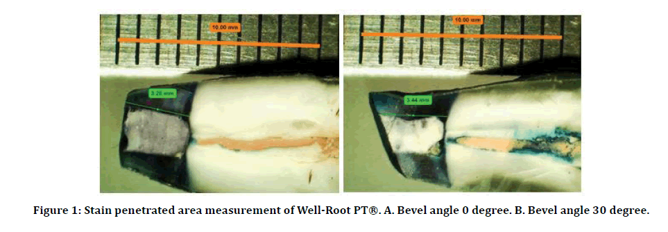
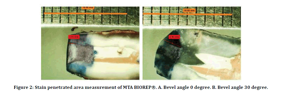
Samples were digitally viewed and photographed using digital camera connected to stereomicroscope (BLS 200, NTB series zoom, Italy) under magnification of 20x and the micrographs transported in a JPEG format to a computer to be saved. Micro leakage measured by utilizing Kinovea software 2018® (Version 2, Boston, USA) to assess the degree of stain penetration in millimeters along the interface between retrograde filling materials and the canal wall. (Figures 1 and 2).
Figure 1: Stain penetrated area measurement of Well-Root PT®. A. Bevel angle 0 degree. B. Bevel angle 30 degree.
Figure 2: Stain penetrated area measurement of MTA BIOREP®. A. Bevel angle 0 degree. B. Bevel angle 30 degree.
Bevel angle 30 degree
The results analyzed statistically by using (SPSS version 21, Chicago, IL, USA) involving Minimum, maximum, mean and standard deviation (SD) as a descriptive statistics. Also, an inferential statistics including (Two Way ANOVA) with Bonferroni test, partial eta square effect size and both D'Agostino and Shapiro normality tests at P<0.05.
Results
Descriptive statistical analysis obtained for stain penetration for each group is presented in Table 1. The results shade lights on that all cavities filled with retrograde filling materials showed microleakge with varying degrees. MTA BIOREP (M) demonstrated highly statistical significant differences compared with Well-Root PT (P) at 0 degree and 30 degree bevel angles as presented in Table 1.
Table 1: Mean, standard deviation, and comparison of the root end filling groups with respect to micro leakage values utilizing a Two Way ANOVA.
| Angles | Fillings | Min. | Max. | Mean | SD | Mean difference | F | P value | Partial eta square Effect size |
|---|---|---|---|---|---|---|---|---|---|
| 0º | P | 2.12 | 4.08 | 3.277 | 0.695 | 2.184 | 84.813 | 0.000 HS | 0.527 Large |
| M | 0.34 | 1.95 | 1.093 | 0.533 | |||||
| 30º | P | 2.57 | 4.09 | 3.457 | 0.447 | 1.8095 | 58.221 | 0.000 HS | 0.434 Large |
| M | 0.26 | 3.67 | 1.647 | 1.133 |
HS=highly significant at p<0.01.
Partial eta square=small (0.01-0.059), medium (0.06-0.139), large (>=0.14)
P value=clinical significance
Effect size=practical significance.
Discussion
The root end filling material must have a perfect sealing ability property to impede leakage of microorganisms and its toxins toward surrounding tissues. The success of endodontic surgery depends mostly on sealing ability of retrograde filling materials. More over studies showed that the degree of microleakage determined by sealing ability of root end filling materials, bevel angle and depth of resection. Various materials have been utilized as a retrograde filling substances and the selection of these substances can be chose according to sealing ability, manipulating properties, biocompatibility and a clinical records of success. Methylene blue stain penetration technique was chosen to evaluate micro leakage due to high degree of staining, easy of manipulation and lower molecular weight than of microorganism’s toxins. In this study, we integrated stain penetration technique and the utilization of Kinovea program for more precise and standardization of micro leakage assessment of apical cavities. According to the conditions of this study, the results exhibited that the two materials showed micro leakage but there was less leakage significantly higher with MTA BIOREP® than with Well Root PT®.
Because of MTA BIOREP ® filling material have almost comparable composition to that of traditional MTA so it’s regarded to have efficient sealing ability and biocompatibility. These characteristics favor its use as a root end filling material which is motivate us to select it as one of the materials tested in this study.
The result of this study comes in agreement with the preceding studies of dye leakage which have been performed on MTA employing various kinds of stains. These outcomes harmonious with prior studies where MTA has been shown to be more desirable than other retrograde filling materials.
The hydrophilic characteristics and the creation of interfacial surface that placed between the dentinal wall and the filling material may allow the MTA BIOREP ® to have greater marginal sealing ability. It was established that further hydration of MTA powder may result in leakage decrease and compressive strength increase. Also the MTA has the ability to precipitate of hydroxyapatite crystals in the moisture presence which may be relevant in leakage decreasing afterwards. Another advantages including excellent adherence to the walls of retrograde cavity and low degree of solubility resulting micro leakage reduction [11-18].
In this study the MTA BIOREP® may show the hydroxyapatite crystals creation at the interface between retrograde cavity wall and filling material as a consequence the substance reveal superb bonding thus hindering the dye penetration and hence presented less micro leakage. Also it’s believed that MTA BIOREP® is hydrophilic so it can undergo expansion during setting when it’s placed in a humid environment that lessens the micro leakage.
Conclusion
Taking into account the restrictions of this study it can be concluded the followings:
The type of filling material and the resection angle influence the extent of apical microleakge.
Correctly prepared and handled MTA BIOREP ® filling material give us excellent results regarding micro leakage.
Declerations
Conflict of Interest the authors declare that there are no potential conflicts of interest related to the study.
Source of Funding
Nil.
Ethical Clearance
This research has exemption as it a routine treatment (no new materials were used).
References
- Sousa CJA, Loyola AM, Versiani MA, et al. A comparative histological evaluation of the biocompatibility of materials used in apical surgery. Int Endod J 2004; 37:738–748.
- Holt GM, Dumsha TC. Leakage of amalgam, composite, and super-EBA, compared with a new retrofill material: bone cement. J Endod 2000; 26:29-31.
- Von Arx T. Apical surgery: A review of current techniques and outcome. Saudi Dent J 2011; 23:9-15.
- Mandava P, Bolla N, Thumu J, et al. Microleakage evaluation around retrograde filling materials prepared using conventional and ultrasonic techniques. J Clin Diagnostic Res 2015; 9:43-46.
- Saini D, Nadig G, Saini R. A comparative analysis of microleakage of three root end filling materials – an in vitro study. Archives Orofac Sci 2008; 3:43-47.
- Ingle JI, Bakland LK. Endodontics. 5th Edn. Baltimore: BC Decker.Elsevier 2002.
- Torabinejad M, Watson TF, Pitt ford TR. Sealing ability of a mineral trioxide aggregate when used as a root end filling material. J Endod 1993; 19:591-595.
- Bondra DL, Harwell GR, MacPherson MG, et al. Leakage in vitro with IRM, high cooper amalgam, EBA cement as retrofilling materials. J Endod 1989; 15:157-160.
- Erbasar GN H, Tulumbaci F, Erbasar R C. Evaluation of microleakage of three different retrograde filling materials in apical resection using an AutoCad program.EU Dishek Fak Derg 2018; 39:98-104.
- Johnson BR. Considerations in the selection of a root-end filling material. Oral Surg Oral Med Oral Pathol Oral Radiol Endod 1999; 87:398-404.
- Camps J, Pashley D. Reliability of the dye penetration studies. J Endod 2003; 29:592-594.
- Torabinejad M, Smith PW, Kettering JD, et al. Comparative investigation of marginal adaptation of mineral trioxide aggregate and other commonly used root end filling materials. J Endod 1995; 21:295-299.
- Aqrabawi J. Sealing ability of amalgam, super EBA cement, and MTA when used as retrograde filling materials. Brit Dent J 2000; 88:266-268.
- Kubo CH, Gomes APM, Mancini MNG. In vitro evaluation of apical sealing in root apex treated with demineralization agents and retrofilled with mineral trioxide aggregate through marginal dye leakage. Braz Dent J 2005; 16:187-191.
- Sarker NK, Caicedo P, Moiseyeva R, et al. Physicochemical basis of the biologic properties of mineral trioxide aggregate. J Endod 2005; 31:97-100.
- Chong BS, Pitt Ford TR, Hudson MB. A prospective clinical study of Mineral Trioxide Aggregate and IRM when used as root-end filling materials in endodontic surgery. Int Endod J 2003; 36: 520–526.
- Perez AL, Spears R, Gutmann JL, et al. Osteoblasts and MG-63 osteosarcoma cells behave differently when in contact with ProRoot MTA and white MTA. Int Endod J 2003; 36:564-570.
- Shipper G, Grossman ES, Potha AG, et al. Marginal adaptation of mineral trioxide aggregate (MTA) compared with amalgam as a root end filling material: a low-vacuum (LV) versus high-vacuum (HV) SEM study. Int Endod J 2004; 37:325-336.
Author Info
Ahmed Abdulkareem Mahmood* and Hassanien Ahmed Al-jumaily
1Department of Oral and Maxillofacial Surgery, College of Dentistry, University of Baghdad, IraqReceived: 06-Mar-2021 Accepted: 29-Mar-2021 Published: 05-Apr-2021


