Case Report - (2022) Volume 10, Issue 11
A Case Series of Avascular Necrosis of Left Hip Managed by Core Decompression with Bone Marrow Injection Procedure
Ruthirakumar M*, Vasanthkumar and Ganesh MT
*Correspondence: Dr. Ruthirakumar M, Department of Orthopaedics, Sree Balaji Medical College and Hospital, Bharath Institute of Higher Education and Research, Chennai, Tamil Nadu, India, Email:
Abstract
Aim: The main aim of the study is to prospectively analyse the functional outcome of avascular necrosis of hip managed by core decompression with bone marrow injection procedure.
Methods: A prospective analysis of 17 patients (9 men and 8 women, with mean age 35 years) with modified ficat and alert classification avascular necrosis of hip managed by core decompression with bone marrow injection procedure between July 2019 and July 2021 was performed. The outcomes were studied based on Range of Motion (ROM) and Visual Analog Score (VAS).
Results: The mean ROM and VAS at the interval of 3 weeks, 5 weeks, 7 weeks, 6 months, 1 year shows excellent improvement. The mean VAS score is 8.8 and range of motion is excellent.
Conclusion: In the present study, 17 patients with avascular necrosis of hip managed by core decompression with bone marrow injection procedure. Management of avascular necrosis of hip by core decompression with bone marrow injection allows for early rehabilitation of the patient and has excellent functional outcome with less incidence of complications. Hence, we strongly recommend considering it in the management of avascular necrosis of hip.
Keywords
Avascular necrosis, Hip, Core decompression, Bone marrow injection, Prospective study
Introduction
Avascular necrosis of hip is the condition in which there is loss of blood supply to the femoral head. Blood supply to the femoral head (ball of hip joint) is interrupted by traumatic and non-traumatic. Avascular necrosis of hip mainly due to traumatic and non-traumatic. Traumatic causes are fracture neck of femur and fracture dislocation of hip injuries. Non-traumatic causes are steroid use, excess alcohol intake, sickle cell disease, other blood cell disorders, deep sea divers and miners [1-2].
Classification
Avascular necrosis of hip can be classified on modified FICAT and alert classification based on radiological investigation (Table 1).
| Stage | Radiological findings |
|---|---|
| I | Plain radiograph, magnetic resonance imaging, and scintigraphy: normal |
| llA | Sclerotic and cystic lesion (absence of subchondral cystic formation) |
| llB | Subchondral collapse (crescent sign) and/or subchondral aliasing |
| Ill | Irregular femoral contour |
| IV | Collapse of the femoral head, acetabula involvement, and articular destruction (osteoarthritis) |
Table 1: Modified FICAT and alert classification.
• Stage 1: X-ray, MRI, Scintigraphy normal
• Stage 2A: Sclerotic and cystic lesion (absence of subchondral cyst)
• Stage 2B: Subchondral collapse (crescent sign)
• Stage 3: Irregular femoral contour
• Stage 4: Collapse of femoral head, acetabular involvement and articular destruction
Aim and objective: The main purpose of the study is to prospectively analyse the functional outcome of avascular necrosis of hip managed by core decompression with bone marrow injection procedure [3-5].
Methods: There are 3 patients with an age group of 25 to 40 years of age. Avascular necrosis of hip was classified on modified FICAT and alert and treatment is then processed. Out of these, 2 patients were avascular necrosis of hip stage 2A. Patient was treated with core decompression with bone marrow injection.
Case Presentation
A prospective analysis of 17 patients (11 men and 6 women, with mean age 35 years) with Modified FICAT and alert classification. Avascular necrosis of hip managed by core decompression with bone marrow injection procedure between July 2019 and July 2021 was performed [6-8].
The patient outcome was studied based on Range of Motion (ROM) and Visual Analog Score (VAS). In this study we included only avascular necrosis of Hip. Informed consent and written consent were taken from all patients. Surgery was done electively after assessment under regional anaesthesia. All cases were taken up for surgery immediately following admission.
Methodology: The management method was decided after classifying avascular necrosis of hip by modified FICAT and alert classification.
The patients were taken for surgery as early as possible time depending on their co-morbidities and skin condition. Avascular necrosis of hip was classified according to modified FICAT and alert classification.
Preoperative preparation: Patient underwent a preoperative evaluation including the following parameters: HB, blood sugar, ECG, renal function test, x ray chest in order to get fitness for surgery.
All surgeries were done under C arm guidance. Fractures were managed by core decompression with bone marrow injection.
Follow up period: At 1 week, 3 weeks, 5 weeks, 1 month, 3 months, and 5 months. K wire removal at the period of 7-9 weeks [9-12].
Results
The mean rom and vas at the interval of 1 week, 3 weeks, 5 weeks, 1 month, 3 months and shows excellent improvement. Mean vas score is 8.8 and range of motion is excellent.
The result shows the efficacy of the functional outcome of avascular necrosis of hip managed by core decompression with bone marrow injection procedure.
Discussion
Patient came with C/O pain in the left hip for past 1 month, aggravated for past 3 days. Patient was unable to weight bear on the left lower limb. Pain was insidious in onset, progressive in nature, pricking type of pain not associated with numbness, aggravated by walking and relieved on rest.
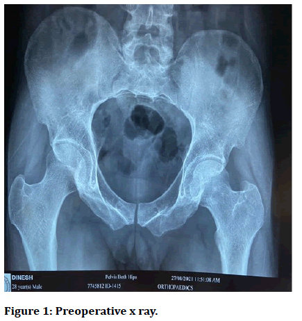
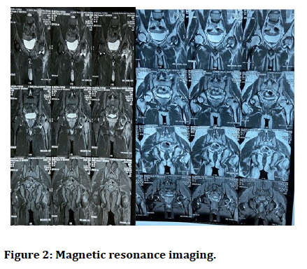
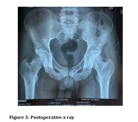
No h/o trauma/injury; no h/o fever, no h/o weight loss, no h/o loss of appetite. Patient is a known alcoholic for past 10 years (3 quarters/day for last 4 years); occasional smoker for past 10 years. Patient went to alcohol de-addiction centre for rehabilitation 2 years back. Bowel and bladder habits normal; normal sleep pattern (Figures 1-3) [12-15].
Plan done: Left hip core decompression with bone marrow injection (under spinal anaesthesia).

Figure 1: Preoperative x ray.

Figure 2: Magnetic resonance imaging.

Figure 3: Postoperative x ray.
The patient was placed in supine position on fracture table, via lateral. Approach, 6 cm incision made on the lateral aspect of the leg at the base of greater trochanter, skin and subcutaneous tissue cut and retracted. Tensor fascia LATA split. Lateral aspect of femur is visualized; bone window made at base of greater trochanter guide wire is passed. Through the bone window to the head of femur (superior and anterior)
Aspect via C arm guidance, reaming is done over guide wire using inner reamer (8 mm) of triple reamer. Bone tissue from the reamed sent for HPE.
Bone marrow aspiration: From the iliac crest 5 cm posterior to ASIS passing bone marrow needle. Aspirated around 5 ml injected into the head of femur via cortical window. Bone wax used to plug cortical window. Thorough wound wash given. Tensor fascia LATA sutured. Skin and subcutaneous tissue closed in layers; sterile dressing done.
After core decompression of left hip, patient symptomatically improved after series follow up with physic and mobilization was done. Core decompression with bone marrow injection is the best modality of treatment for avascular necrosis of left hip stage 2A. Then Patient symptomatically improved, Suture removal was done and then patient discharged and asked for regular follow up [16,17].
Conclusion
Treatment of avascular necrosis of hip is very important or else it may lead to many complications like hip collapse, subchondral fracture and early osteoarthritis. This study is done in such a way that all the patients treated well with the treatment of choice and patient was symptomatically improved. After this patient mobilized well and started doing regular activities.
References
- FICAT RP. Idiopathic bone necrosis of the femoral head. Early diagnosis and treatment. J Bone Joint Surg 1985; 67:3.
- Lafforgue P, Dahan E, Chagnaud C, et al. Early stage avascular necrosis of the femoral head: MR imaging for prognosis in 31 cases with at least 2 years of follow up. Radiol 1993; 187:199.
- Stulberg BN, Bauer TW, Belhobek GH. Making core decompression work. Clin Orthop Relat Res 1990; 186.
- Aaron RK, Lennox D, Bunce GE, et al. The conservative treatment of osteonecrosis of the femoral head. A comparison of core decompression and pulsing electromagnetic fields. Clin Orthop Relat Res 1989; 209.
- Kane SM, Ward WA, Jordan LC, et al. Vascularized fibular grafting compared with core decompression in the treatment of femoral head osteonecrosis. Orthopedics 1996; 19:869.
- Markel DC, Miskovsky C, Sculco TP, et al. Core decompression for osteonecrosis of the femoral head. Clin Orthop Relat Res 1996; 226.
- Chang MC, Chen TH, Lo WH. Core decompression in treating ischemic necrosis of the femoral head. Zhonghua Yi Xue Za Zhi Chin Med J 1997; 60:130.
- Scully SP, Aaron RK, Urbaniak JR. Survival analysis of hips treated with core decompression or vascularized fibular grafting because of avascular necrosis. J Bone Joint Surg Am 1998; 80:1270.
- Powell ET, Lanzer WL, Mankey MG. Core decompression for early osteonecrosis of the hip in high risk patients. Clin Orthop Relat Res 1997; 181.
- Ha YC, Jung WH, Kim JR, et al. Prediction of collapse in femoral head osteonecrosis: A modified Kerboul method with use of magnetic resonance images. J Bone Joint Surg Am 2006; 88:35.
- Aigner N, Schneider W, Eberl V, et al. Core decompression in early stages of femoral head osteonecrosis an MRI controlled study. Int Orthop 2002; 26:31.
- Herndon JH, Aufranc OE. Avascular necrosis of the femoral head in the adult. A review of its incidence in a variety of conditions. Clin Orthop Relat Res 1972; 86:43–62.
- Mwale F, Wang H, Johnson AJ, et al. Abnormal vascular endothelial growth factor expression in mesenchymal stem cells from both osteonecrotic and osteoarthritic hips. Bull NYU Hosp Jt Dis 2011; 69:S56–S61.
- Lavernia CJ, Sierra RJ, Grieco FR. Osteonecrosis of the femoral head. J Am Acad Orthop Surg 1999; 7:250–261.
- Gangji V, Hauzeur JP. Treating osteonecrosis with autologous bone marrow cells. Skeletal Radiol 2010; 39:209–211.
- Hernigou P, Bachir D, Galacteros F. The natural history of symptomatic osteonecrosis in adults with sickle cell disease. J Bone Joint Surg Am 2003; 85-A:500–504.
- Hernigou P, Poignard A, Nogier A, et al. Fate of very small asymptomatic stage-I osteonecrotic lesions of the hip. J Bone Joint Surg Am 2004; 86-A:2589–2593.
Author Info
Ruthirakumar M*, Vasanthkumar and Ganesh MT
Department of Orthopaedics, Sree Balaji Medical College and Hospital, Bharath Institute of Higher Education and Research, Chennai, Tamil Nadu, IndiaCitation: Ruthirakumar M, Vasanthkumar, Ganesh MT, A Case Series of Avascular Necrosis of Left Hip Managed by Core Decompression with Bone Marrow Injection Procedure, J Res Med Dent Sci, 2022, 10 (11): 178-181.
Received: 12-Sep-2022, Manuscript No. JRMDS-22-57345; , Pre QC No. JRMDS-22-57345(PQ); Editor assigned: 14-Sep-2022, Pre QC No. JRMDS-22-57345(PQ); Reviewed: 28-Oct-2022, QC No. JRMDS-22-57345; Revised: 15-Nov-2022, Manuscript No. JRMDS-22-57345(R); Published: 28-Nov-2022
